^

X-rays (X-ray studies)

Pelvic X-rays in women, men and children

X-rays of the pelvic bones and hip joints can be performed in three projections: anteroposterior (AP), posteroanterior (PA) and lateral.

X-ray of the Turkish saddle: what it is done for, what it shows

X-ray analysis of the sella turcica is a relatively common diagnostic procedure, including in gynecology.

X-ray of the clavicle in two projections

In clinical practice, X-ray imaging remains one of the leading methods of instrumental diagnostics for injuries and diseases of the musculoskeletal system.

X-rays of the ribs

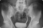
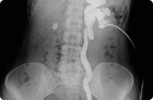
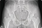
When X-raying the ribs, the condition of the bone mechanism is visualized, and the spine can be partially seen. The degree of ionizing radiation is not considered dangerous to human health, so X-rays can be considered a good alternative to ultrasound examination.

X-ray of the scapula

Since it is not always possible to immediately identify the cause of the pathology, doctors use an X-ray of the scapula. This is a non-invasive, painless and accessible diagnostic method, which is also quite informative.

Digital X-ray

What is this new diagnostic method – digital X-ray? In fact, it is a familiar X-ray examination with obtaining an image processed digitally.

X-ray of the cervical spine with functional tests

The need for an X-ray of the cervical spine is assessed by the doctor in each specific situation. In most cases, this diagnostic method allows the medical specialist to quickly and accurately determine the diagnosis and begin adequate treatment.

Pyelography

One of the radiological methods for diagnosing diseases of the urinary system is pyelography (pyeloureterography, ureteropyelography), in which the examination of the kidneys and ureters is carried out using special contrast agents.

X-ray of the coccyx

X-ray of the coccyx is an informative diagnostic method that helps to identify many bone and joint pathologies in the corresponding area of the spinal column.