
All iLive content is medically reviewed or fact checked to ensure as much factual accuracy as possible.
We have strict sourcing guidelines and only link to reputable media sites, academic research institutions and, whenever possible, medically peer reviewed studies. Note that the numbers in parentheses ([1], [2], etc.) are clickable links to these studies.
If you feel that any of our content is inaccurate, out-of-date, or otherwise questionable, please select it and press Ctrl + Enter.
X-ray of the Turkish saddle: what it is done for, what it shows
Medical expert of the article

Last reviewed: 04.07.2025
Today, medicine uses many methods for diagnosing diseases and injuries. One of the long-standing proven methods for many years has been radiography, which provides valuable information to doctors of various specialties. Often, an X-ray of the sella turcica is prescribed - a deep bone formation in the sphenoid bone of a person, in which the pituitary gland is located - an endocrine gland. The study is prescribed for various pathological disorders of a neuroendocrine, neurological or neuro-ophthalmic nature. [ 1 ]
Indications for the procedure
The sella turcica is a bony element, so its condition can be monitored using X-ray examination. The results of such diagnostics are often necessary in neuropathological, ophthalmological and gynecological practice. [ 2 ]
The bony bed of the pituitary gland in the form of the sella turcica is examined in the following situations:
- for the diagnosis of neuroendocrine disorders, for the detection of tumor processes affecting the hypothalamic-pituitary mechanism;
- with elevated levels of the hormone prolactin in the blood;
- with increased intracranial pressure;
- in case of abnormal development of the skull;
- in case of growth deficiency or its pathological acceleration;
- in case of traumatic brain injury;
- for menstrual cycle disorders in women and girls;
- for visual disorders of unclear etiology;
- in case of infertility (after hormonal studies);
- for regular headaches.
Why do they do an X-ray of the sella turcica?
X-ray diagnostics helps to identify tumor processes in the pituitary gland (for example, prolactinoma ), osteoporosis, changes in the vascular network due to increased intracranial pressure. The image demonstrates the configuration, outlines and dimensions of the bone element being examined. If its dimensions are increased, the entrance to it is widened, a double contour appears, then it is possible to assume the development of a tumor in the pituitary gland and competently prescribe subsequent diagnostics.
X-ray of the sella turcica is prescribed by a doctor to confirm or refute various pathologies. Most often, patients are referred for examination if there is a suspicion of a pituitary tumor.
X-ray of the sella turcica in gynecology
X-ray analysis of the sella turcica is a relatively common diagnostic procedure, including in gynecology. There are a number of indications for a gynecologist to prescribe this type of examination:
- severe menstrual dysfunction of unknown origin;
- inability to conceive, infertility;
- increased amounts of prolactin in the blood (prolactinemia).
Pituitary disorders in female patients often manifest themselves as systematic poor health, deterioration of skin turgor, early appearance of wrinkles. Hair suffers: hair becomes brittle and falls out. Digestive disorders are often a concern, appetite decreases, constipation is observed. To understand the causes of such manifestations, the doctor may need additional diagnostics, including a gynecological examination with smear cytology, laboratory hormonal studies, pelvic ultrasound, and sometimes magnetic resonance imaging. A comprehensive approach to diagnostics allows you to establish the cause of the disorder and prescribe an effective therapeutic regimen.
Preparation
There is no need for any special preparation of the patient to perform an X-ray of the sella turcica.
Immediately before the diagnostic procedure, the patient should remove all metal accessories and jewelry from the head and neck area - that is, earrings, glasses, chains, hairpins, etc. If there are prostheses - for example, a cochlear implant - then the doctor should be warned about this in advance. Removable dentures and hearing aids should be removed. It is advisable to unbutton the top buttons of clothing.
There are no restrictions on food and drink consumption or medication intake.
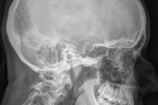
Technique a turkish saddle x-ray
X-rays of the sella turcica can be either general or targeted. During general radiography, the doctor examines the condition of the cranium as a whole. Targeted radiography involves a more thorough examination of individual parts of the skull – in particular, the sella turcica.
During the imaging procedure, the patient may be sitting, standing, or lying on their back or stomach, depending on the desired image projection. The radiologist will usually take 1-2 images. The most common position is:
- the patient lies on his back, arms are placed along the body, chin is lowered;
- the head is turned at an angle of 45 degrees to the side being examined (you can put a soft cushion underneath for convenience);
- the head is fixed with an elastic bandage;
- Nearby organs are covered with protective lead plates.
During the X-ray fixation process, the patient should not breathe or swallow.
Another way of laying:
- the patient lies on his stomach or sits, with his head pressed sideways against the surface of the table;
- the upper limbs are located along the body, the forearms rest on the surface of the table;
- the shoulder and chin adjacent to the table should be slightly raised using a wedge-shaped roller so that the median sagittal cranial plane is parallel to the plane of the X-ray film;
- the head is fixed.
After the diagnostic procedure is completed, the patient is sent home or to the doctor, depending on the situation.
X-ray of pathology of the sella turcica
X-rays of the sella turcica may reveal pituitary tumors. Signs of such a pathological process are:
- local or total areas of osteoporosis, characterized by thinning of the bone;
- atrophic changes in the bone walls;
- unevenness of the inner contour of the saddle;
- thinning of the sphenoid processes;
- the emergence of a "double contour".
The listed signs indicate the presence of a small pituitary tumor. At the same time, the doctor may also pay attention to such pathological manifestations as thickening of the occipital and frontal bones, calcification of the dura mater of the brain and the formation of calcifications in the brain tissue.
A large sella turcica on an X-ray may indicate such pathological conditions as a pituitary adenoma, cyst, aneurysm, primary hypothyroidism, increased intracranial pressure, etc. If necessary to confirm or clarify the diagnosis, the doctor may additionally prescribe a computer or magnetic resonance imaging. Modern tomographic equipment helps to detect even very small neoplasms.
In some patients, X-rays reveal the so-called "empty sella turcica". In the image, the pathology is located under the diaphragm of the bone formation and is manifested by the following signs:
- symmetry of the bottom in the frontal plane;
- vertical increase in formation, closed configuration;
- double contour of the bottom on the sagittal image.
Symptoms of such changes may be absent, therefore, when an "empty sella turcica" is detected, patients are often only followed dynamically. Disorders of the pituitary function are more typical for women.
Contraindications to the procedure
Every person in everyday life receives a certain dose of radiation from natural radiation sources. This dose is approximately equal to 1 μSv, as it depends on many factors - in particular, on the place of residence, on working conditions, etc.
In situations where it is necessary to conduct a reliable diagnosis to prescribe subsequent effective treatment, the potential harm from radiation may not be taken into account, since the benefit from diagnostics becomes much more important. Although some contraindications do exist and are considered relative: if the benefit exceeds the potential harm, then the diagnostic study is still carried out. Such contraindications may include:
- decompensated cardiovascular diseases;
- pregnancy period (especially the first trimester);
- extreme exhaustion of the patient;
- early childhood.
The decision on the possibility and necessity of conducting an X-ray of the sella turcica is made by the attending physician.
Normal performance
The resulting X-ray image of the sella turcica – a lateral craniogram – is carefully studied by a doctor, who evaluates the contours of the bone formation, its size and configuration, and the condition of the element as a whole.
The following diagnostic indicators are considered normal:
- the normal sagittal index is 9-15 mm;
- the normal vertical indicator is 7-12 mm;
- the value of the ratio of the height and length of the saddle, the so-called saddle index - in children more than 1, in older patients less than 1.
Complications after the procedure
X-ray is a very common diagnostic method for both adults and children. Although children are prescribed the examination only if there are vital indications. If the risk of complications from the pathological process exceeds the possible harm from the radiation dose of X-ray, doctors perform diagnostics, giving preference to modern X-ray equipment.
In general, the destructive effect of X-rays largely depends on the duration and degree of radiation exposure, and the frequency of imaging. If the irradiation is prolonged, then the risk of developing certain complications cannot be ruled out:
- blood diseases;
- cataracts, visual impairment;
- oncological processes, benign tumors;
- metabolic disorders;
- premature aging;
- disorders in the functioning of the reproductive system.
Doctors insist: a single diagnostic test within the recommended limits cannot lead to consequences of this kind. In addition, the power of the X-ray machine, its type, and its regime equipment play an important role. It has been proven that modern digital devices do not lead to any negative effects even if they are used repeatedly.
Consequences after the procedure
X-ray of the sella turcica, like other types of X-ray examination, is associated with a fairly high radiation load. However, modern diagnostic devices, the operation of which is based on digital technologies, have a much smaller volume of ionizing radiation, unlike outdated technology. Therefore, it is safe to say that X-ray has become a fairly safe procedure today. Although it is also not worth abusing the study.
Doctors insist that this type of diagnostics does not pose any particular danger. The moment of the X-ray image and the exit of the rays from the emitter lasts only a fraction of a second. At the same time, non-aggressive, filtered radiation is emitted.
To reduce the likelihood of adverse effects, X-rays of the sella turcica are not recommended for pregnant patients or small children without good indications.
Care after the procedure
No special care is required for the patient after an X-ray of the sella turcica. However, if a person wants to protect themselves from additional radiation exposure, it is better to take care of this in advance. It is advisable that the following products predominate in the diet:
- nuts (walnuts, almonds);
- oatmeal, beans, lentils;
- apples, pears;
- pumpkin, squash;
- seaweed, seafood.
After receiving any dose of radiation, you should eat more fiber to help remove radioactive decay products from the body.
In addition, you need to:
- drink clean water, up to 2-3 liters per day for an adult;
- eat more vegetables, greens, berries and fruits;
- add dairy products to your diet, especially cottage cheese and sour cream.
In addition to water, you can drink dried fruit infusion, green tea, fresh juices (only natural, self-pressed). You can drink some dry wine (about 50 ml).
It is necessary to walk a lot in the fresh air, lead an active lifestyle, and then an X-ray of the sella turcica will not leave any adverse effects on the body.