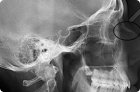
There are several methods of performing X-rays: without the use of a contrast agent (survey image) and with its use, allowing monitoring of its movement inside the kidneys, ureter, and bladder.
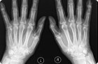
X-ray of hands does not require preliminary preparation. The only requirement is the absence of metal objects on them: rings, bracelets. If there is a plaster cast at the time of the X-ray, it is removed.
The most accessible, informative and painless method of visualizing bone structures is radiography. The image also clearly shows damage to joints, cartilage of traumatic and inflammatory genesis, congenital defects.
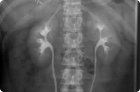
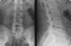
The most accessible diagnostic method that allows visualization of the internal structure and assessment of the condition of the skeletal bones of the spine is radiography.
Radiography is a method of radiation diagnostics and is a non-invasive study of the internal structure of a certain part of the body by shining X-rays through it and obtaining a projection of the image on a special film.
During the procedure, several images are taken simultaneously. Everything is determined by the range of motion in the joint being examined. The method used is called double contrast.
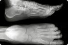
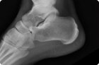
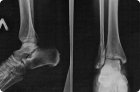
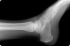
Nowadays, traumatology increasingly deals with various injuries. One of the weakest organs that is most often subject to injury is the foot. It is quite easy to damage it.
The most widely used non-invasive diagnostic method for detecting congenital and acquired pathological changes in bone and joint tissue is visualization of their anatomy using X-rays.
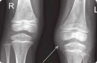
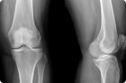
Knee pain, joint mobility impairment in this area and traumatic injuries are quite common reasons for visiting a doctor. Even an experienced doctor cannot easily determine by eye what the unpleasant symptoms are associated with.