
All iLive content is medically reviewed or fact checked to ensure as much factual accuracy as possible.
We have strict sourcing guidelines and only link to reputable media sites, academic research institutions and, whenever possible, medically peer reviewed studies. Note that the numbers in parentheses ([1], [2], etc.) are clickable links to these studies.
If you feel that any of our content is inaccurate, out-of-date, or otherwise questionable, please select it and press Ctrl + Enter.
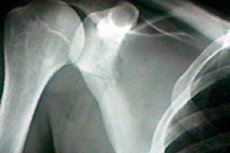
X-ray of the scapula
Medical expert of the article

Last reviewed: 06.07.2025
The scapula is one of the bones of the human musculoskeletal system. It has a triangular shape and connects the humerus and the collarbone. When this anatomical structure is damaged, sharp pain appears and mobility is limited. Since it is not always possible to immediately identify the cause of the pathology, doctors use an X-ray of the scapula. This is a non-invasive, painless and accessible diagnostic method, which is also quite informative. [ 1 ]
Indications for the procedure
X-ray of the scapula is a mandatory examination method if there is a suspicion of a fracture of this bone, as well as for assessing the dynamics of bone fusion after a violation of integrity. Among other possible indications:
- assumption of the presence of a tumor process, benign or malignant;
- infectious and inflammatory foci in the scapular region.
An X-ray of the scapula may be required:
- if the patient indicates pain in the scapular region;
- if there is a dislocation of the shoulder joint;
- if there is a suspicion of a cystic formation or bursitis;
- if the mobility of the shoulder joint is limited.
When receiving an X-ray of the scapula, the doctor has the opportunity to:
- learn the features of the location of the scapula in relation to adjacent joints and humerus bones;
- identify changes in interarticular gap dimensions, monitor the condition of the tendon and cartilage apparatus;
- examine bone structures, diagnose partial and complete fractures, dislocations of the scapula;
- detect areas of tissue necrosis.
Preparation
An X-ray of the scapula does not require any special preparation. It is advisable to refrain from smoking and drinking alcohol.
There is no need to change your diet or stick to any special diet, but it is better to refrain from eating or drinking for several hours before the examination. It is best to take an X-ray of the scapula on an empty stomach.
When going to the procedure, it is necessary to wear clothes without complicated fasteners, loose, which can be easily and quickly removed (the patient will have to undress to the waist). Any metal jewelry and accessories that may get on the image and interfere with visualization should be left at home. [ 2 ]
Positioning the patient for x-ray of the scapula
To obtain an anteroposterior projection image, the patient stands upright, with his back and shoulder blade resting against a vertical post. The opposite side is not pressed against the post, but is moved away from it by 15 degrees. The shoulder blade being examined is parallel to the bar. The patient lifts the chin, turns the head to the side opposite to the examination. The arm on the diagnostic side is raised and fixed on the back of the head, or brought to the hip and bent. X-ray radiation is directed perpendicular to the film, from front to back. Central radiation is directed to the center of the shoulder blade and film. You should hold your breath after exhaling. The position is correct if there is a free zone between the ribs and the shoulder blade, the medial and lateral scapular edges are aligned, and the shoulder blade is completely visualized. [ 3 ]
To obtain a lateral projection image, the technique differs from the previous one: the patient stands at a vertical stand and presses the necessary side against it. The upper limb from the side being examined is placed on the head or on the thigh. The opposite shoulder is slightly moved to the side (the arm is held in front), the lateral and medial edges of the scapula are combined. The directed X-ray radiation goes along a tangent line relative to the scapula and perpendicular to the film. The center is directed to the middle of the scapula (in the middle of the axillary fossa). Breathing is held. [ 4 ]
Another, less common option for laying (for traumatic cases):
- the patient stands with his back or lies on his back, the side being examined is abducted at an angle of 45 degrees, the upper limbs are bent at the elbows and are on the stomach;
- the center is directed through the shoulder and the area between the shoulder blade and the ribs, along the level of the armpit and further into the center of the cassette.
Contraindications to the procedure
A regular X-ray of the scapula has virtually no contraindications due to the risk of adverse effects of X-rays on the human body. Contraindications are relative, which means the following: if diagnostics can save the patient's life, then it is performed in any case.
When is it not recommended to perform a scapula x-ray?
- For women during pregnancy and lactation.
- For children in the absence of compelling indications (up to 14-15 years old).
- For patients with decompensated conditions.
- Patients who have already received large amounts of radiation in the last few months.
- Insufficiency of renal and hepatic function.
- Severe thyroid pathologies.
- Individual sensitivity to X-ray radiation.
Normal performance
Usually, the X-ray image can show the consequences of the disorder, such as trauma. These can be chips, complete or partial fractures. It is also possible to see signs of an inflammatory reaction, the presence of seals, congenital defects (in particular, changes in the configuration and size of the scapula). [ 5 ]
A scapula fracture is determined on an X-ray by a change in the color of the bone and the presence of a clear darkening line. With such damage, it is important to identify the type of fracture:
- fracture of the scapular neck;
- body and angles of the scapula;
- scapular articular process;
- scapular spine;
- coracoid and acromial process of the scapula.
Scapula fractures are relatively rare, occurring in approximately 1-2% of all bone fractures. They may occur after a fall on the back, due to direct impact. More often, a transverse fracture of the body of the scapula is noted on X-ray, in the area below the spine, and somewhat less often - a fracture of the neck and processes. In isolated cases, longitudinal damage to the body of the bone is found, which is accompanied by a strong divergence of the fragments. [ 6 ]
When examining a scapular neck fracture on an x-ray, a radiologist can distinguish between a single fracture and a multi-fragment fracture. A multi-fragment fracture is said to occur when there are one or more completely separated intermediate bone fragments.
The suprahumeral and coracoid process are often broken off by direct blows to the scapula area, by falling on the back from a great height or with support on the upper limb. A fracture of the coracoid process of the scapula on an X-ray may be combined with rib injuries.
X-ray anatomy of the scapula
When deciphering an X-ray image, traumatologists and orthopedists need to know the anatomical features and be able to spatially construct anatomical elements with an indication of the change in their relationship to each other, which is expressed in degrees and millimeters.
The scapula is a kind of triangle adjacent to the back surface of the chest in the space from the second to the seventh rib. Taking into account the shape of the bone, three edges are distinguished:
- medial edge (“looks” at the spine);
- lateral edge;
- the upper edge where the scapular notch is located.
The specified edges are connected at certain angles. One of these angles, the lower one, is directed downwards, and the upper and lateral ones are located at the ends of the upper scapular edge. The lateral angle is thicker than the others and has a slightly deepened glenoid cavity. The edge of the cavity is separated from the rest of the scapula by the neck.
Above the upper border of the acetabulum there is an elevation, a tubercle, to which the tendon of the long head of the biceps brachii is attached. The lower border also has a similar elevation with the attachment of the long head of the triceps brachii. From the upper border of the scapula near the glenoid cavity, the coracoid process extends. [ 7 ]
The anterior, or parcostal, surface of the scapula is a flattened depression called the subscapular fossa. The spine of the scapula runs along the posterior plane, dividing this surface into two depressions: the supraspinous and infraspinous fossae. [ 8 ]
The scapula from the posterior projection is a triangular formation with three edges, angles and processes. At the base of the coracoid process, a notch can be seen: inexperienced specialists may mistake it for an area of bone destruction, which is especially common during the diagnosis of elderly patients with signs of senile calcification, when the notch is transformed into a hole.
Complications after the procedure
Many injuries and pathologies of the scapula cannot be accurately diagnosed without an X-ray. Accordingly, it becomes difficult to choose the appropriate treatment. Visual examination only allows us to assume a particular disorder, so in many cases an X-ray is simply necessary.
During the procedure using a modern digital device, the patient receives minimal radiation exposure. Even when taking 2-3 images, no harm is done to the body.
But it is highly undesirable to perform an X-ray examination on women during pregnancy – especially in the first trimester. However, in exceptional cases (for example, in case of a fracture or for diagnosing serious pathologies), such a method is indispensable. In order to protect the future baby, the doctor uses protective shielding plates and aprons covering the patient’s stomach and chest during the X-ray. If the situation allows, it is better to opt for X-ray rather than computed tomography.
Consequences after the procedure
X-rays have the ability to break down molecules, so their influence can theoretically lead to the destruction of cell membranes and damage to nucleic acids DNA and RNA. But the theory and the real danger are somewhat different. Experts say that modern digital X-ray machines give out a lower radiation dose than old diagnostic devices. Each X-ray examination and dose must be recorded by the doctor in a special dose load log. The entry is also made in the patient's outpatient card. [ 9 ]
The effective dose of X-rays is measured in mSv or μSv. Modern X-ray machines are equipped with a built-in dosimeter that determines the amount of radiation received by the patient. Such a dose, even with a similar study, can be different, which depends on the area of the body, and the distance to the X-ray tube, etc.
An X-ray of the scapula is considered a safe diagnostic. A person receives a much greater radiation load during computed tomography and fluoroscopy:
- Fluoroscopy takes several minutes, while an X-ray takes a fraction of a second;
- During a computed tomography scan, a series of images are taken, and the more there are, the higher the radiation load.
The likelihood of harm to the body can be reduced by using special protective equipment: lead pads, plates, shields.
It is not recommended to do several types of X-ray examinations in 1-2 days: the body needs recovery after the diagnostic procedure.
Care after the procedure
No special care measures are required after X-ray examination. However, to level out the radiation load received, specialists recommend:
- after the procedure, you should drink a lot of water, tea, compotes, and also get a good night's sleep (cell regeneration occurs mainly at night);
- at least for a while, it is necessary to give up bad habits, not to smoke and not to drink alcohol (drinking a small amount of dry wine is allowed);
- physical activity and walks in the fresh air are encouraged: aerobic exercise reduces the risk of developing cancer;
- It is advisable to at least temporarily give up fatty foods, fast food, sweets, smoked foods, and carbonated drinks.
A diet that includes plant foods in your diet will be beneficial:
- cabbage (broccoli, white cabbage);
- grape;
- beet;
- pomegranate;
- blueberries, raspberries, currants;
- seaweed.
The consumption of dairy products, nuts, oatmeal, buckwheat, and prunes is encouraged.
If everything is done correctly, the patient's body recovers within 24 hours after a scapula X-ray. During this period of time, radioactive substances completely disintegrate and are eliminated.