
All iLive content is medically reviewed or fact checked to ensure as much factual accuracy as possible.
We have strict sourcing guidelines and only link to reputable media sites, academic research institutions and, whenever possible, medically peer reviewed studies. Note that the numbers in parentheses ([1], [2], etc.) are clickable links to these studies.
If you feel that any of our content is inaccurate, out-of-date, or otherwise questionable, please select it and press Ctrl + Enter.
Bladder X-ray for adults and children
Medical expert of the article

Last reviewed: 06.07.2025
Bladder X-ray is one of the most common medical procedures. However, it requires preliminary preparation. It has its own indications and contraindications for implementation.
As a rule, any X-ray examination of the urinary system begins with a survey X-ray. The kidneys and upper urinary tract are subject to examination. It will be necessary to prepare for the procedure in advance. In particular, in the evening, on the eve of the examination, it is necessary to carry out a preliminary cleansing enema. In the morning on the day of the examination itself, you can allow yourself a light breakfast. If an X-ray examination of the bladder is to be carried out, one cleansing enema in the morning, directly on the day of the procedure, is often enough. If the procedure is planned for young people who have a fairly well-functioning intestine, in particular, there are no problems with its cleansing, an X-ray examination may not be required. [ 1 ]
The procedure itself is as follows: first, an image of the kidney area is taken, then the ureter and bladder are examined. The procedure allows you to evaluate the shape, position of the kidneys, their functional and anatomical features, as well as the specific structure of the surrounding bone skeleton and muscles. The edge of the lumbar muscle is also quite clearly visible on the overview image. This makes it possible to assess its condition and exclude pain irradiation in the presence of acute or chronic pain syndrome. The overview procedure allows you to assess the general condition of the body, evaluate the features of the location and functioning of the genitourinary tract. It is also possible to promptly detect stones in the kidneys and ureters, study the structural and functional features of the bladder. In men, the prostate gland and urethra are subject to additional examination. Oxalates, phosphates and carbonates are especially well visualized in overview radiography, since they very well retain X-ray radiation. The surrounding tissues are much less able to retain X-ray radiation, so the presence of stones contrasts sharply with the background of the surrounding tissues. Urate, xanthine or cystine stones may be detected as a faint shadow.
Sometimes, plain radiography can diagnose phleboliths. This is an inflammatory-degenerative disease of the veins of the urogenital tract. It occurs mainly in the pelvic cavity. In this case, individual sections of the veins are subject to calcification. In some cases, nearby lymph nodes may be subject to calcification, which happens if there are areas of neoplasms. In order to confirm or refute the presence of a neoplasm, additional research methods are used. In particular, it is possible to visualize stones and separate them from shadows using a plain radiograph, which is taken in a direct or oblique projection. In this case, a catheter is inserted into the ureter. If there is a stone in the kidneys or ureter, its shadow completely coincides with the shadow of the catheter in both projections. If the shadow comes from phleboliths, lymph nodes, neoplasms, it is located separately from the catheter, often turning in the opposite direction.
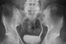
Cystography
Cystography is one of the types of X-ray examination of the genitourinary tract. The procedure is performed by filling the bladder with a sergosin solution. If it is not available or cannot be administered, gas (oxygen) is used. Accordingly, an X-ray image is obtained, which shows the cavity of the bladder. The nature of the image can be used to judge the presence or absence of pathological changes in the genitourinary tract. Thus, normally the bladder is completely filled with a contrast agent and has a rounded shape. At the same time, the density of the contrast agent is the same, its contours are smooth.
Using the cystography method and its various modifications, it is possible to detect stone shadows, including urate shadows. Thus, urates have the appearance of radiolucent areas. Using cystography, it is possible to establish a differential diagnosis in many pathological conditions. In particular, it is possible to subtly differentiate the localization of a urinary stone. Thus, if the stone is located in the bladder or in the lower segment of the ureter, this will be clearly visible on an X-ray. The procedure allows for a differential diagnosis in both urology and gynecology. In particular, using this method, it is possible to distinguish the shadow of a calcified myomatous node affecting the uterus from the shadow of a stone or neoplasm (node) located in the genitourinary tract. It is possible to diagnose a bladder calculus. Cystography is often used to diagnose diverticula (volvulus) of the bladder, to identify abnormalities in its development. An important role is given to the diagnosis of the tumor process. In this case, it is possible to easily diagnose both benign and malignant neoplasms. In addition, it is possible to assess the size, localization features, size, degree of infiltration of the tumor process, and the walls of the bladder. [ 2 ]
Cystography plays an important role in diagnosing tuberculous lesions of the bladder, in the development of viral or bacterial infections. This is especially important if it is not possible to catheterize the bladder. Cystography can be used to determine uretecel, accurately determine its size, location, and other important characteristics. This method is indispensable in diagnosing bladder hernias, in determining the degree of bladder deviation, which is extremely important when performing plastic surgery. In particular, this method is used both immediately before and immediately after the bladder plastic surgery procedure. In gynecology, this method allows you to identify the presence of a connection between diseases of the bladder and uterus, as well as its appendages, to determine the degree of their mutual influence on the course of the pathological process, on the development of dysuric pathologies and disorders. The method is used to differentiate conditions such as pericystitis, paracystitis, and other forms of cystitis. This method can be used to recognize various forms of bladder fistulas, diagnose various forms of reflux. It is also possible to diagnose neurogenic forms of bladder disease.
The cystography method is irreplaceable in diagnosing congenital and acquired anomalies of the urinary bladder. First of all, we are talking about such conditions as bladder exstrophy, anomalies of the apex of the urinary bladder, urachuses, and double urinary bladder.
In the presence of a double bladder, this is clearly visible on the image. Thus, the bladder is divided into two independent parts by a partition. In this case, each cavity has a separate connection with the urethra. That is, the urethra is separate for each part of the urethra... in fact. And the image visualizes a double urethra. Or one of the halves of the bladder opens into the urethra. Cystogram is the basis for diagnosis. In this case, additional research methods are often not required. When analyzing the images, you can notice that they clearly show two halves of the bladder. Between them, a partition is clearly visible. In the apex area, this partition is represented by an oval contour. A shadow appears that resembles a heart of cards. Also, sometimes with the help of a cystogram, it is possible to diagnose bladder anomalies that appear in the form of an hourglass. In this case, one half of the bladder is located directly above the other. In this case, the study is carried out in the craniocaudal direction.
Urography of the bladder
Urography of the urinary bladder is a diagnostic procedure in which a solution of a monoatomic, diatomic, or triatomic iodine compound (sergosin, diodone, or triiotrast, respectively) is injected into a vein. The molecules of these substances are excreted by the kidneys. In this case, free iodine is not released. Accordingly, a phenomenon known as iodism occurs, resulting in contrasting of the urinary tract. The contrast agent completely fills the renal pelvis, is excreted through the ureter, and penetrates the urinary bladder. A series of images is taken (at certain intervals). In this case, all sections of the urinary tract are examined. The first image is taken 7-10 minutes after the administration of the contrast agent, the second image is taken approximately 15-20 minutes later, and the third - 30-40 minutes after the administration of the contrast agent. [ 3 ]
The procedure has a number of advantages, in particular, it is an absolutely painless method. It is non-invasive, the risk of injury is completely excluded. Preliminary catheterization of the bladder and cystoscopy are not required. Another advantage of the method is that it is possible to examine the morphological picture of the urinary tract, as well as study their structural and functional state, examine the structural and functional features of the urinary tract, kidneys (both one and both at the same time). However, it is worth noting that the clarity of the image sometimes leaves much to be desired, in particular, it is significantly inferior to the methods of retrograde pyelography. It is especially difficult to conduct research using this method if the kidney function is reduced.
It is also necessary to take into account that the procedure has some contraindications. In particular, the procedure cannot be performed in acute liver diseases, in many blood diseases and hematopoietic dysfunction, in Graves' disease, and also during menstruation. A strict contraindication is high azotemia.
Indications for the procedure
The main indications for X-ray examination of the bladder are pathology of the kidneys and urinary system. The procedure is carried out in case of structural, functional disorders of the kidneys, ureters, urogenital bladder, in the presence of acute and chronic inflammatory processes, in case of suspected development of a tumor process, traumatic injury, congenital anomalies of the urogenital tract. The presence of diverticula, altered function of the urinary system, can serve as a direct indication for X-ray examination of the urogenital tract. It is worth noting that this procedure can be used for both men and women, and even for children. It is noteworthy that the procedure can be carried out for urological and gynecological diseases and suspicions of them, has an important diagnostic value. The procedure plays an important role in differential diagnostics. [ 4 ]
The procedure is performed when an atypical process develops in the bladder. This can be inflammation, cystic formations, tumors). If there are stones, sand, other formations and foreign bodies in the bladder, this procedure is also performed. Indications for the procedure are also the presence of acquired and congenital anomalies of the bladder, urinary incontinence of various origins, the presence of enterovisical fistulas. It is often prescribed in the presence of complications of various origins that occur after an infectious or inflammatory process. Indications include diagnoses such as urethritis, cystitis, urolithiasis, and suspicion of these pathologies. It is also performed in case of injuries, when planning surgical operations, after them. [ 5 ]
Preparation
The procedure is quite simple and does not require serious preparation. However, it is necessary to follow some recommendations. This will make the procedure as informative, accurate and effective as possible. So, a few days before the procedure, you need to exclude the use of certain products, in particular, those that lead to intense gas formation. You should exclude coffee, strong tea, carbonated drinks, cabbage, beans, peas, and other legumes. Dairy products are contraindicated. Immediately before the manipulation, a cleansing enema is performed, or you can take laxatives. If the excretory function of the intestine is good, you can do without an enema.
Technique bladder X-rays
To perform the procedure, the patient is asked to take a horizontal position. Then a sterile catheter is inserted into the bladder cavity. With its help, approximately 200-250 ml of fluid is introduced. The technique for further performing the procedure is quite simple. After the bladder is filled with a contrast agent, the examination begins. Pictures are taken. They are taken in different directions, in several projections. This allows for an image to be taken in several positions. In particular, the image is taken in the supine position, lying on the side. The procedure is performed at the moment of urination, and immediately after that. Then the catheter is removed, and a control picture is taken (an image of an empty bladder is taken). [ 6 ]
A descending method of performing the procedure is also possible, in which the contrast agent is injected into a vein. Then, after about 40-60 minutes, the procedure is performed. However, this method is not very convenient and is characterized by a certain degree of pain. If necessary, anesthesia is used. [ 7 ]
X-ray of the kidneys and bladder
One of the most common procedures is an X-ray of the kidneys and bladder. The procedure is performed in the presence of inflammatory and infectious processes, as well as in the diagnosis of anomalies of the kidneys and bladder. The procedure is indicated if diverticulosis, bladder exstrophy are suspected. In case of bladder exstrophy, the first thing that attracts attention is the absence of the symphysis on the X-ray. This occurs due to the divergence of the pubic bones. The bones diverge by approximately 8-12 cm. The anterior pelvic semiring remains underdeveloped. Other anomalies are often visualized, primarily affecting the skeletal system. Also, the pathological process is often accompanied by abnormal development of the kidneys and upper urinary tract.
X-ray of the kidneys and bladder is also the only method for the final diagnosis of bladder diverticula. First of all, it is worth noting that diverticula can be congenital or acquired. Both conditions can be detected using cystography. Often the former are called true, and the latter - false. This is due to the fact that false diverticula are often a consequence of the development of stagnation, and are formed in patients with various disorders of urine outflow from the bladder. Also, this disease often occurs against the background of the prostate, with difficulty urinating. A contrast agent is used to perform an X-ray. Thus, when diagnosing, it is necessary to take into account that when a true diverticulum is formed, a muscular sphincter is formed at the site of its connection with the bladder. With rapid administration of a contrast agent, as well as in the case when an unheated substance is used, a clamping of the muscular sphincter may occur, which will complicate further administration of the contrast and make the procedure impossible or difficult. Therefore, if true diverticulosis is suspected, a heated contrast agent should be used. Its temperature should not be lower than body temperature. The substance should also be administered slowly, in small quantities (no more than 150 ml).
X-ray of the bladder with contrast agent
When diagnosing bladder diseases, an X-ray of the bladder is often prescribed. The procedure is performed using an endoscope. It is performed if there are indications. It can be performed at any age. It allows you to evaluate the main parameters of the bladder. For this, a contrast agent is poured into it (in the form of a special solution). First of all, with the help of this method, you can evaluate the structural features of the organ, as well as its integrity. It allows you to promptly identify pathology and begin timely treatment. This procedure is usually prescribed by a urologist, less often by a surgeon.
There are two methods of performing the procedure: ascending and descending. In the ascending method, the contrast is injected into the bladder using a catheter. The total amount of contrast agent is 150-200 ml. In the second case, with the descending method of performing the procedure, intravenous administration of contrast is used. It takes about 45 minutes for the substance to reach the ureter. Several types of contrast agents are widely used, in particular, triombrast, urografin, iodamine. X-ray allows you to quickly identify pathologies of the bladder. It is especially important to use this method in case of reflux, cystitis, fistulas, in the presence of neoplasms, diverticula, stones, anomalies of the genitourinary tract and kidneys. [ 8 ]
This method allows diagnosing various forms of urinary incontinence, as well as assessing the excretory function of the kidneys. This is done approximately 30 minutes after the procedure.
X-ray of the bladder of a child
Sometimes it is necessary to conduct an X-ray of the bladder of a child. The procedure is not performed on newborns. It is performed no earlier than 5 months. Doctors often use this method when absolutely necessary, since irradiation or administration of a contrast agent in childhood is undesirable. However, sometimes there is no other way. In this case, you need to prepare for the procedure in advance. So, about a week before the procedure, the child should be on a diet. You should exclude foods that cause gas incontinence, bloating. On the day of the procedure, a cleansing enema is performed. This allows you to achieve bowel cleansing. Basically, a medicinal enema is performed, using anti-inflammatory drugs. This helps to avoid inflammation. Before the procedure, a drug test is performed. [ 9 ]
Indications for the procedure in children include conditions such as inflammation, infectious processes, the presence of structural and functional changes in the kidneys and genitourinary tract.
The technique is as follows: first, the required amount of contrast agent is injected through the urethra. The contrast agent is injected before and after the discharge. Children under one year of age, as well as restless children, are given anesthesia. You must not drink or eat for 6 hours before the procedure. The procedure lasts approximately 15 minutes. However, after the procedure, the child must be under medical supervision for 2 hours. During this time, the child must empty the bladder so that the contrast agent completely leaves the body. It will take approximately 24 hours for complete excretion. During this time, you need to drink plenty of fluids.
Contraindications to the procedure
The procedure has certain contraindications for use, for example, it cannot be performed in cases of recent bladder surgery. In case of bladder obstruction, the procedure is also not recommended. A strict contraindication is the presence of an acute inflammatory process in acute form. The procedure is not performed during pregnancy, especially in the early stages. In case of a strong allergic reaction and intolerance to contrast agents, this procedure is also not performed. The procedure is contraindicated in acute liver and kidney failure.
Complications after the procedure
The procedure has virtually no consequences. For example, after the procedure, increased thirst may be observed for some time, especially in children. In the first 24 hours after the procedure, plenty of fluids are required, since the contrast agent is being excreted. Some people may have intolerance to the contrast agent, so a drug test should be performed in advance. Children often undergo the procedure under anesthesia, so medical observation is required for 2-3 hours. [ 10 ]
As a rule, there are no complications after the bladder X-ray procedure. Thus, the only possible complication is an allergic reaction to the contrast agent. Therefore, in order to avoid complications, it is necessary to conduct a drug test in advance.
Care after the procedure
Bladder X-ray is a relatively simple procedure that does not cause complications and does not require special care after the procedure. Adults can resume their normal daily routine immediately after the procedure. Children should be under medical supervision for 2-3 hours. This is due to the fact that the procedure is performed on children mainly with the use of a contrast agent and anesthesia, so it is necessary to monitor the child's reaction until the contrast and anesthesia are completely eliminated. Within 24 hours after the procedure, you need to drink plenty of fluids, which will speed up the elimination of the contrast.