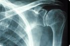
Radiography is a diagnostic procedure with a solid history, already more than 120 years old. But despite the development of new modern methods of diagnosing various diseases, it has not lost its relevance to this day.
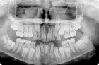
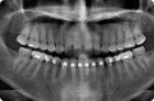
Used in dental and maxillofacial radiology, an orthopantomogram (OPG) is a panoramic X-ray image of the upper and lower jaws, teeth, craniofacial bones and joints, maxillary sinuses and adjacent areas.
If a person has a toothache, he rushes to the dentist for help and insists on treatment, not to remove such a treasure. But the dentist is not God, he cannot see the condition of the diseased tooth from the inside.
Among the instrumental examination methods in dentistry, dental orthopedics, and maxillofacial surgery, the most informative is a panoramic x-ray of the jaw.
The essence of the manipulation consists of introducing a contrast fluid into the cavity of the vessel while simultaneously taking a series of X-ray images.
X-ray of abdominal organs - radiography - is a traditional diagnostic method of clinical medicine based on localized irradiation with a minimal dose of X-rays, which results in projection images of the internal structures of the body.
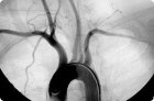
Coronary angiography continues to be the “gold standard” for diagnosing coronary artery stenosis, determining the effectiveness of drug therapy, PCI and CABG.
Contrast ventriculography (VG) is one of the important catheterization angiographic methods. Ventriculography is the contrasting of the ventricle of the heart with recording of the image on film or another recording device (video film, computer hard or CD-disk).