
All iLive content is medically reviewed or fact checked to ensure as much factual accuracy as possible.
We have strict sourcing guidelines and only link to reputable media sites, academic research institutions and, whenever possible, medically peer reviewed studies. Note that the numbers in parentheses ([1], [2], etc.) are clickable links to these studies.
If you feel that any of our content is inaccurate, out-of-date, or otherwise questionable, please select it and press Ctrl + Enter.
Pyelography
Medical expert of the article

Last reviewed: 03.07.2025
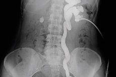
One of the radiological methods for diagnosing diseases of the urinary system is pyelography (pyeloureterography, ureteropyelography), in which the examination of the kidneys and ureters is carried out using special contrast agents. [ 1 ]
Indications for the procedure
When examining the kidneys, the need for pyelography is determined by the doctor, especially if patients complain of intense pain in the kidney area, problems with urination, and also in the presence of hematuria (blood in the urine). And when other visualization methods do not allow to determine the condition of such structures as the renal pelvis (Pelvis renalis), cups (Calices renales) and ureters (Ureter), they resort to pyelography - an X-ray of the kidneys with the introduction of a contrast agent. [ 2 ]
Dysfunction of the listed structures of the urine accumulation and excretion system is possible with various pathologies and diseases of the kidneys, and the task of diagnostics is to find their possible causes. In addition, pyelography can be used to identify developmental anomalies of the kidneys (hyper- and hypoplasia, medullary sponge kidney, diverticula of the renal calyces, etc.) and to check the location of a catheter or ureteral stent. [ 3 ]
For better visualization (image enhancement), iodine-containing water-soluble non-ionic contrast agents are used for pyelography, for example, Iopamidol, Pamirei, Optirey, Ultravist 300, etc. [ 4 ]
Preparation
Preparation for this kidney examination includes stopping (several days before) taking analgesics, neuroleptics, antidepressants, beta-blockers; in the evening before the procedure – stopping eating after 6-7 pm and cleansing the intestines with a laxative.
On the day of the examination, in the morning you also do not eat food (or drink liquids) and cleanse your intestines again by doing an enema.
At the medical facility, you should change into loose home clothes, remove jewelry and any metal objects that may interfere with obtaining X-ray images.
Who to contact?
Technique pyelography
In pyelography, the technique used depends only on the method by which the radiopaque substance is administered.
Retrograde pyelography or ascending pyelography involves the introduction of a contrast agent into the orifice of the corresponding ureter through the urethra using a cystoscope through which a catheter is inserted, and through it the contrast agent. The procedure requires epidural anesthesia. [ 5 ]
Antegrade pyelography, which is more often used when upper urinary tract obstruction is suspected, is performed by introducing a contrast agent through a skin puncture (needle puncture) into the lateral area of the back - directly into the renal pelvis. In this case, the accuracy of the puncture and the movement of the injected drug from the kidney to the ureter and bladder are monitored fluoroscopically. The procedure is anesthetized with a local anesthetic. [ 6 ]
Minimally invasive intravenous pyelography or excretory pyelography is also performed, in which a contrast agent is injected into a vein in the arm at regular intervals. The procedure is monitored and controlled using continuous fluoroscopy, which converts X-rays into video images. [ 7 ]
A series of X-ray images (photos are also taken at intervals) and video, which is produced by an X-ray machine and a detector (located above the patient lying motionless on the table) allow an assessment of the conductivity of the ureters and urinary tract, which may be impaired due to the presence of kidney stones, tumors, congenital anomalies, and in men - due to hyperplasia or tumor of the prostate gland. [ 8 ]
Contraindications to the procedure
Pyelography is contraindicated in pregnancy, elevated body temperature, exacerbation of any existing diseases, allergy to iodine, hyperthyroidism and thyrotoxicosis, acute or chronic renal failure (including chronic diabetic nephropathy), malignant blood diseases.
Relative contraindications include diabetes mellitus, severe arterial hypertension, decreased circulating blood volume (hypovolemia), and old age (over 70 years).
Complications after the procedure
Due to the use of radiocontrast agents containing iodine, negative consequences of pyelography are possible in the form of deterioration of kidney function (with a decrease in the glomerular filtration rate and an increase in the level of creatinine in the blood serum), convulsions, tachycardia, shortness of breath, and the development of anaphylactic shock.
Possible complications of retrograde pyelography: nausea and/or vomiting, pain during urination, bleeding, urinary tract infections, sepsis. And with antegrade pyelography there is also a risk of formation of a urinary cyst.
Care after the procedure
The type of pyelography performed determines what care is required for patients and how long their rehabilitation will be after the procedure. In an outpatient setting or in a ward of a medical institution where the patient is undergoing inpatient treatment, medical staff must monitor his condition: monitor heart rate, breathing, blood pressure. Also, diuresis and the presence of blood in the urine are monitored during the day (a small amount of blood immediately after antegrade or ascending pyelography is considered normal).
If urination is painful, the doctor will prescribe painkillers that do not reduce blood clotting.
If at home after pyelography a fever develops; the puncture site becomes red, wet or painful; the amount of blood in the urine increases or urination becomes difficult, you should immediately contact your doctor.
Reviews
Reviews of specialists in the medical literature about this method of visualizing the structures of the urinary system indicate that today, in many cases, ultrasound examination is used - ultrasound of the kidneys and ureters (including with color Doppler mapping), computed [ 9 ] or magnetic resonance imaging.