Examination of the body

Microbiologic and bacterioscopic examination of vaginal discharge

Microbiological and bacterioscopic examination is used to diagnose inflammatory processes and allows to establish the state of the vaginal biocenosis, as well as some pathogens of sexually transmitted diseases. This examination is carried out during a woman's initial visit to a gynecologist, as well as before gynecological operations and diagnostic manipulations.

Mammary gland (breast) examination

Examination and palpation of the mammary glands by a gynecologist is as necessary as examination of the cervix in mirrors during a gynecological examination. During the examination, it is necessary to pay attention to the structure of the mammary glands, their size (hypoplasia, hypertrophy, graphic changes).

Bimanual vaginal examination

Vaginal (internal) examination is performed with the middle and index fingers of one hand (usually the right). The other hand must first spread the labia. Vaginal examination allows you to determine the condition of the pelvic floor muscles, large glands of the vestibule, urethra, vagina

Methods of research of the autonomic nervous system

When studying the autonomic nervous system, it is important to determine its functional state. The principles of the study should be based on a clinical and experimental approach, the essence of which is functional-dynamic studies of tone, autonomic reactivity, and autonomic support of activity.

Thermal imaging (thermography)

Thermography is the registration of invisible infrared radiation. The maximum radiation is at a wavelength of 9.5 microns. According to the Stefan-Boltzmann law, the amount of emitted energy is proportional to the fourth power of the absolute temperature: W=T4.

Electroencephalography in epilepsy

The definition of an epileptic seizure through the concept of neuronal discharge determines the most important significance of EEG in epileptology. Clarification of the form of epilepsy (more than 50 variants) includes as a mandatory component a description of the EEG pattern characteristic of this form. The value of EEG is determined by the fact that epileptic discharges, and therefore epileptiform activity, are observed on EEG outside of an epileptic seizure.

Electroencephalogram disorders in diseases

Tumors of the cerebral hemispheres cause slow waves to appear on the EEG. When the median structures are involved, bilateral synchronous disturbances may join local changes. A progressive increase in the severity of changes with tumor growth is typical. Extracerebral benign tumors cause less severe disturbances.

Computer methods for analyzing electroencephalograms

The main methods of computer analysis of EEG used in the clinic include spectral analysis using the fast Fourier transform algorithm, instantaneous amplitude mapping, spikes, and determination of the three-dimensional localization of the equivalent dipole in brain space.

Deciphering electroencephalography results

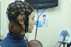
EEG analysis is performed during the recording and finally upon its completion. During the recording, the presence of artifacts (induction of network current fields, mechanical artifacts of electrode movement, electromyogram, electrocardiogram, etc.) is assessed, and measures are taken to eliminate them. The frequency and amplitude of the EEG are assessed, characteristic graphic elements are identified, and their spatial and temporal distribution is determined.

Methods of electroencephalography

In common practice, EEG is recorded using electrodes placed on intact scalp surfaces. Electrical potentials are amplified and recorded. Electroencephalographs have 16-24 or more identical amplification and recording units (channels) that allow simultaneous recording of electrical activity from the corresponding number of pairs of electrodes placed on the patient's head.