Examination of the body

Corneal sensitivity study

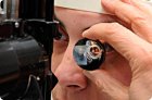
The cornea is a highly sensitive membrane of the eyeball. In various pathological conditions of the eye, its sensitivity can significantly decrease or completely disappear, so its determination can be a very informative indicator when establishing a diagnosis.

Intraocular pressure study

The level of intraocular pressure can be determined in various ways: approximately (by palpation), using applanation or impression type tonometers, and also in a non-contact manner.

Gonioscopy

Gonioscopy is a method of examining the angle of the anterior chamber, hidden behind the translucent part of the cornea (limbus), which is performed using a gonioscope and a slit lamp.

Biomicroscopy

Biomicroscopy is intravital microscopy of eye tissues, a method that allows one to examine the anterior and posterior sections of the eyeball under different lighting conditions and image sizes.

Ophthalmoscopy

Ophthalmoscopy is a method of examining the retina, optic nerve and choroid in rays of light reflected from the fundus. The clinic uses two methods of ophthalmoscopy - in reverse and direct form.

Examination of the eye under lateral (focal) and transmitted illumination

The method is designed to detect subtle changes in the anterior part of the eyeball. The study is conducted in a dark room using a table lamp installed to the left and in front of the patient at a distance of 40-50 cm at the level of his face.

Methods of accommodation research

It is necessary to distinguish between absolute and relative accommodation. Absolute accommodation is the accommodation of one (isolated) eye when the other is excluded from the act of vision.

Methods of refraction research

The most common subjective method of refraction testing is the method based on determining the maximum visual acuity with correction.

Skiascopy

Skiascopy (from the Greek scia - shadow, scopeo - I examine) is a method of objectively studying clinical refraction, based on observing the movement of shadows obtained in the area of the pupil when the latter is illuminated using various techniques.

Visual acuity: Visual acuity test

To study visual acuity, special tables are used containing optotypes of different sizes (letters, numbers, signs).