
All iLive content is medically reviewed or fact checked to ensure as much factual accuracy as possible.
We have strict sourcing guidelines and only link to reputable media sites, academic research institutions and, whenever possible, medically peer reviewed studies. Note that the numbers in parentheses ([1], [2], etc.) are clickable links to these studies.
If you feel that any of our content is inaccurate, out-of-date, or otherwise questionable, please select it and press Ctrl + Enter.
Ophthalmoscopy
Medical expert of the article

Last reviewed: 04.07.2025
Ophthalmoscopy is a method of examining the retina, optic nerve and choroid in rays of light reflected from the fundus. The clinic uses two methods of ophthalmoscopy - in reverse and direct form. Ophthalmoscopy is more convenient to perform with a wide pupil.
The pupil is not dilated if glaucoma is suspected, so as not to cause an attack of increased intraocular pressure, as well as in case of atrophy of the sphincter of the pupil, since in this case the pupil will remain dilated forever.
Reverse ophthalmoscopy
It is intended for a quick examination of all sections of the fundus. It is carried out in a darkened room - an examination room. The light source is installed to the left and slightly behind the patient. The ophthalmologist stands opposite the patient, holding an ophthalmoscope in his right hand, placed against his right eye, and sends a light beam into the eye being examined. An ophthalmic lens with a power of +13.0 or +20.0 D, which the doctor holds with the thumb and index finger of his left hand, is installed in front of the eye being examined at a distance equal to the focal length of the lens - 7-8 or 5 cm, respectively. The patient's other eye remains open and looks in the direction past the doctor's right eye. The rays reflected from the patient's fundus hit the lens, are refracted on its surface and form on the doctor's side in front of the lens, at its focal length (respectively 7-8 or 5 cm), a real, but 4-6 times magnified and inverted image of the examined areas of the fundus hanging in the air. Everything that seems to be lying on top actually corresponds to the lower part of the examined area, and what is outside corresponds to the internal areas of the fundus.
In recent years, aspherical lenses have been used in ophthalmoscopy, which allows obtaining a virtually uniform and highly illuminated image across the entire field of view. The image size depends on the optical power of the lens used and the refraction of the eye being examined: the greater the lens power, the greater the magnification and the smaller the visible area of the fundus, and the magnification in the case of using the same lens power when examining a hypermetropic eye will be greater than when examining a myopic eye (due to the different lengths of the eyeball).
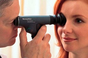
Direct ophthalmoscopy
Allows you to directly examine the details of the fundus revealed by reverse ophthalmoscopy. This method can be compared to examining objects through a magnifying glass. The examination is performed using mono- or binocular electric ophthalmoscopes of various models and designs, allowing you to see the fundus in a direct view magnified 13-16 times. In this case, the doctor moves as close as possible to the patient's eye and examines the fundus through the pupil (preferably against the background of drug-induced mydriasis): the patient's right eye with the right eye, and the left eye with the left eye.
With any method of ophthalmoscopy, examination of the fundus is carried out in a certain sequence: first, the optic nerve head is examined, then the area of the yellow spot (macular area), and then the peripheral parts of the retina.
When examining the optic disc in reverse, the patient should look past the doctor's right ear if the right eye is being examined, and at the examiner's left ear if the left eye is being examined. Normally, the optic disc is round or slightly oval, yellowish-pink in color, with clear boundaries at the level of the retina. Due to the intensive blood supply, the inner half of the optic disc has a more saturated color. In the center of the disc there is a depression (physiological excavation), this is the place where the optic nerve fibers bend from the retina to the cribriform plate.
The central retinal artery enters through the central part of the disc and the central retinal vein exits. The central retinal artery in the area of the optic nerve disc is divided into two branches - the upper and lower, each of which in turn is divided into the temporal and nasal. The veins completely repeat the course of the arteries. The ratio of the diameter of the arteries and veins in the corresponding trunks is 2:3. The veins are always wider and darker than the arteries. During ophthalmoscopy, a light reflex is visible around the artery.
Outside the optic nerve, at a distance of two disk diameters from it, there is a yellow spot, or macular area (anatomical area of central vision). The doctor sees it during examination, when the patient looks directly into the ophthalmoscope. The yellow spot has the appearance of a horizontally located oval, slightly darker than the retina. In young people, this area of the retina is bordered by a light strip - the macular reflex. The central pit of the yellow spot, which has an even darker color, corresponds to the foveal reflex. The picture of the fundus in different people differs in color and pattern, which is determined by the saturation of the retinal epithelium with pigment and the content of melanin in the vascular membrane. With direct ophthalmoscopy, there are no light glare reflections from the retina, which facilitates the examination. The head of the ophthalmoscope has a set of optical lenses that allow you to clearly focus the image.
Read also: Confocal scanning laser ophthalmoscopy
Ophthalmochromoscopy
The method was developed by Professor A. M. Vodovozov in the 60-80s. The examination is carried out using a special electric ophthalmoscope, which contains light filters that allow the fundus to be examined in purple, blue, yellow, green and orange light. Ophthalmochromoscopy is similar to direct ophthalmoscopy, it significantly expands the doctor's capabilities when establishing a diagnosis, and allows the earliest changes in the eye to be seen that are not visible in normal lighting. For example, the central area of the retina is clearly visible in red-free light, while small hemorrhages are clearly visible in yellow-green light.