
All iLive content is medically reviewed or fact checked to ensure as much factual accuracy as possible.
We have strict sourcing guidelines and only link to reputable media sites, academic research institutions and, whenever possible, medically peer reviewed studies. Note that the numbers in parentheses ([1], [2], etc.) are clickable links to these studies.
If you feel that any of our content is inaccurate, out-of-date, or otherwise questionable, please select it and press Ctrl + Enter.
Methods of refraction research
Medical expert of the article

Last reviewed: 07.07.2025
The most common subjective method of refraction examination is the method based on determining the maximum visual acuity with correction. Ophthalmological examination of the patient, regardless of the suspected diagnosis, begins with the use of this diagnostic test. In this case, two tasks are consistently solved: determining the type of clinical refraction and assessing the degree (magnitude) of clinical refraction.
Maximum visual acuity should be understood as the level achieved with correct, complete correction of ametropia. With adequate correction of ametropia, maximum visual acuity should approach the so-called normal and designated as complete, or corresponding to "one". It should be remembered that sometimes, due to the peculiarities of the structure of the retina, "normal" visual acuity can be greater than 1.0 and be 1.25; 1.5 and even 2.0.
Methodology of implementation
To conduct the study, a so-called spectacle frame, a set of trial lenses and test objects for assessing visual acuity are required. The essence of the method is to determine the effect of trial lenses on visual acuity, while the optical power of the lens (or those - in case of astigmatism) that will provide maximum visual acuity will correspond to the clinical refraction of the eye. The basic rules for conducting the study can be formulated as follows.
- With visual acuity equal to 1.0, it is possible to assume the presence of emmetropic, hypermetropic (compensated by accommodation tension) and weak myopic refraction. Despite the fact that most textbooks recommend starting the examination by applying a lens of +0.5 D to the eye, it is advisable to first use a lens of -0.5 D. With emmetropia and hypermetropia, such a lens under cycloplegia will cause deterioration of vision, and under natural conditions, visual acuity may remain unchanged due to compensation of the power of this lens by accommodation tension. With weak myopia, regardless of the state of accommodation, an increase in visual acuity may be noted. At the next stage of the examination, a lens of +0.5 D should be placed in the trial frame. In case of emmetropia, a decrease in visual acuity will be noted in any case; in case of hypermetropia, an improvement will be observed in conditions of switched off accommodation; and in case of preserved accommodation, vision may remain unchanged, since the lens compensates only part of the latent hypermetropia.
- If visual acuity is less than 1.0, myopia, hyperopia and astigmatism may be assumed. The examination should begin with applying a -0.5 D lens to the eye. In myopia, a tendency towards increased visual acuity will be noted, while in other cases, vision will either deteriorate or remain unchanged. At the next stage, using a +0.5 D lens will reveal hypermetropic refraction (vision will either remain unchanged or, as a rule, improve). If there is no tendency towards a change in visual acuity against the background of correction with spherical lenses, astigmatism may be assumed. To clarify the diagnosis, it is necessary to use special lenses from the trial set - the so-called cylinders, in which only one of the sections is optically active (it is located at an angle of 90° to the cylinder axis indicated on the astigmatic lens). It should be noted that precise subjective determination of the type and especially the degree of astigmatism is a rather labor-intensive process (despite the fact that special tests and methods have been proposed for this purpose). In such cases, the results of objective refraction studies should serve as the basis for establishing a diagnosis.
- After establishing the type of clinical refraction, the degree of ametropia is determined, and by changing lenses, maximum visual acuity is achieved. When determining the magnitude (degree) of ametropia, the following basic rule is followed: from several lenses that equally affect visual acuity, with myopic refraction, the lens with the lowest absolute power is selected, and with hypermetropic refraction, the lens with the highest.
It should be noted that a trial contact correction with a rigid contact lens, which corrects not only ametropia but also aberrations of the anterior corneal surface, can be used to determine maximum visual acuity. In outpatient settings, it is recommended to perform a test with a diaphragm instead of this test. In this case, during the subjective refraction study, visual acuity is determined with trial spectacle lenses and a 2.0 mm diameter diaphragm, which are simultaneously placed in a trial frame. However, the described method has a number of drawbacks that are difficult to eliminate. Firstly, during the study, it is necessary to focus on the level of visual acuity, the decrease of which can be caused not only by the presence of ametropia, but also by pathological changes in the optical media and the neuroreceptor apparatus. In addition, the method is not applicable in the absence of contact with the patient (for example, in young children), as well as simulation and aggravation. In these cases, objective methods of refraction research are more informative, in particular skiascopy, conventional and automatic refractometry, and ophthalmometry.
More accurate data on clinical refraction can be obtained using special devices - refractometers. In a simplified form, the operating principle of these devices can be presented as the registration of light signals reflected from the retina, the focusing of which depends on the type and degree of clinical refraction.
In conventional refractometers (Hartinger, Rodenstock), the adjustment, setting of the required position and type of test mark of the device is carried out manually. In recent years, these devices are practically not used in the clinic.
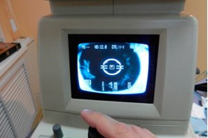
More advanced in terms of objectification of the study are automatic refractometers, in which the analysis of the infrared light beam reflected from the retina is carried out automatically using a special electronic unit. The features of the refraction study technique on these devices are described in detail in the instructions for each of them. The main thing is that refraction studies on automatic refractometers are usually carried out by mid-level medical personnel, and the results are issued as a printout on a special form according to the following main parameters: the value of spherical ametropia, the value of astigmatism, the position of one of the main meridians. Despite the relative high cost of automatic refractometers, in recent years they have gradually become an integral part of the standard equipment of an ophthalmologist's office.
A common drawback of refractometers of various types is the so-called instrumental accommodation, a phenomenon due to which the data obtained during the study may have a shift towards myopic refraction. The reason for this is the impulse to the accommodation tension, caused by the location of the optical part of the device at a small distance from the eye being examined. In some cases, cycloplegia is required to objectify the refractometric data. The latest models of automatic refractometers are equipped with devices that reduce the possibility of instrumental accommodation.
The methods described above are intended to determine the clinical refraction of the eye.
Ophthalmometry
According to foreign terminology, keratometry is an objective method for studying only corneal refraction. The essence of the method is to measure mirror images projected onto the cornea by test marks of the device (ophthalmometer), the dimensions of which, other things being equal, depend on the radius of curvature of the anterior surface of the cornea. During the study, the position of the main meridians of the cornea (in degrees) is determined, as well as the optical power (in diopters) and the radius of curvature of the anterior surface of the cornea (in milliliters) in the specified meridians. It should be noted that there is a clear relationship between the latter indicators: the smaller the radius of curvature of the cornea, the greater its optical power.
Some models of automatic refractometers have a unit with which, during the course of the study, in parallel with clinical refraction (i.e. the general refraction of the eye), corneal refraction is also assessed.
Although the results of ophthalmometry cannot be used to judge the clinical refraction of the eye as a whole, in a number of situations they can be of important and even fundamental significance.
- In diagnostics of astigmatism, the results of ophthalmometry can be used as a starting point. In any case, they should be clarified, if possible, by refractometry and necessarily by subjective examination of refraction. The latter circumstance is related to the possible influence of crystalline astigmatism on the parameters of general astigmatism.
- The data obtained during ophthalmometry (in particular, on corneal refraction), along with the length of the anteroposterior axis, are used in various formulas used to calculate the parameters of refractive surgeries (for example, radial keratotomy) and the optical power of intraocular lenses (IOLs) used to correct ametropia of various origins (for example, hyperopia, which usually occurs after cataract removal ).
- Accurate determination of the radius of curvature of the anterior corneal surface is necessary when choosing such an important parameter of contact lenses as the base radius of their posterior (facing the eye) surface. This measurement is necessary, relatively speaking, to achieve congruence of the anterior corneal surface and the posterior surface of the contact lens.
- The information content of ophthalmometry is quite high in cases of irregular corneal astigmatism, which is usually acquired - formed as a result of various lesions of the cornea (traumatic, inflammatory, dystrophic, etc.). In this case, during the study, a significant increase or, conversely, weakening of the refraction of the cornea, a violation of the mutually perpendicular arrangement of its main meridians, and a distortion of the shape of the mirror image of test marks on the cornea can be detected.
Ophthalmometry can be used to study corneal refraction only in the central zone (2.5-3 mm in diameter). However, even in the absence of astigmatism, the shape of the entire corneal surface differs from spherical and can be geometrically represented as a paraboloid of revolution. In practical terms, this means that even within one meridian, the radius of curvature of the cornea changes: it gradually increases in the direction from the center to the periphery of the cornea, while the refraction of the cornea decreases accordingly. Knowledge of the corneal parameters in the paracentral and even peripheral areas is necessary in a number of clinical situations: when choosing contact lenses and keratorefractive surgeries, determining the degree of influence of various corneal diseases on its refractive properties, etc.
Keratotopographic methods for studying the refraction of the entire surface of the cornea
Research methods that involve assessing the curvature and refraction of the entire surface of the cornea are called keratotopographic, since they can be used to obtain an idea of the relationship between the refraction of different areas of the cornea (conventionally, topography).
An approximate assessment of the refraction of the entire corneal surface can be made using such a simple method as keratoscopy, during which an image of concentrically arranged circles is projected onto the cornea using a simple device (keratoscope). The keratoscope is a disk with illuminated alternating white and black concentric circles. If the cornea has a shape close to spherical, the image is formed from regularly arranged circles. With astigmatism, these images take the form of an oval, and with irregular astigmatism, their orderly arrangement is disrupted. Using a keratoscope, one can only obtain a qualitative assessment of the sphericity of the cornea.
Photokeratographic examination
Photokeratographic examination of corneal topography involves mathematical processing of photokeratograms (pictures of mirror images of circles). In addition, refraction measurement of various corneal areas can be performed using a conventional ophthalmometer equipped with a special attachment for changing the fixation of the patient's gaze (the so-called fixation holometry).
However, the most informative method of studying corneal refraction is computer keratotopography. Special devices (keratotopographs) provide the ability to conduct a detailed objective analysis of refraction and curvature in various areas of the cornea. Keratotopographs have several computer programs for processing the results of the study. A particularly visual option for processing data is also provided using so-called color mapping: the color and intensity of the coloring of various areas of the cornea depends on the refraction of the latter.
The question of the sequence of application of subjective and objective methods of refraction research is important. It is obvious that with the availability of automatic refractometers, objective refractometry can precede the subjective assessment of refraction. However, it is precisely subjective tests that should be of fundamental importance not only in establishing the final diagnosis, but also in choosing an adequate method of correcting ametropia.