Examination of the body

Electroretinography

Electroretinography is a method of recording the total bioelectrical activity of all neurons in the retina: the negative a-wave of photoreceptors and the positive b-wave of hyper- and depolarizing bipolars and Müller cells.

Corneal pachymetry

Pachymetry is a measurement of the thickness of the cornea. It is an indirect sign of the integrity of the corneal endothelium. The greatest thickness of the cornea is at the limbus (0.7-0.9 mm).

Corneal scraping and biopsy

Corneal scraping is performed with a Kimura spatula, a curved needle tip (for the hypodermis) or a blade. After instillation of a local anesthetic without preservatives, the edges and bottom of the lesion (usually an ulcer) are carefully and thoroughly scraped under slit lamp control.

Corneal keratometry

Corneal keratometry is the measurement of the curvature of the axial meridians of the anterior surface of the cornea.

Corneal sensitivity assessment

Assessment of corneal sensitivity in order to identify gross disturbances of corneal sensitivity.

Confocal lifetime microscopy of the cornea

Confocal microscopy of the cornea is one of the modern research methods; it allows for intravital monitoring of the cornea with tissue visualization at the cellular and microstructural level.

Corneal biomicroscopy

Systematic identification of clinical signs, determination of the location, depth and size of corneal damage is of great importance.

Conjunctival examination

The conjunctiva is easily accessible for examination and diagnosis of many of its diseases and does not require any special equipment.

Examination of the lacrimal organs

The marginal tear duct of both eyes is examined with a slit lamp before other lid manipulations or instillation of topical medications that may alter the clinical picture. Many patients with lacrimation do not have significant lacrimation, but on examination they have a high marginal tear duct on the affected side.

Hemodynamic study of the eye

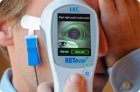
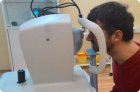
The study of eye hemodynamics is important in the diagnosis of various local and general vascular pathological conditions. The following main methods are used to conduct the study: ophthalmodynamometry, ophthalmoplethysmography, ophthalmosphygmography, rheoophthalmography, ultrasound Dopplerography.