
All iLive content is medically reviewed or fact checked to ensure as much factual accuracy as possible.
We have strict sourcing guidelines and only link to reputable media sites, academic research institutions and, whenever possible, medically peer reviewed studies. Note that the numbers in parentheses ([1], [2], etc.) are clickable links to these studies.
If you feel that any of our content is inaccurate, out-of-date, or otherwise questionable, please select it and press Ctrl + Enter.
Panoramic image of the upper jaw, lower jaw
Medical expert of the article

Last reviewed: 06.07.2025
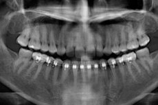
Among the instrumental examination methods in dentistry, dental orthopedics, and maxillofacial surgery, the most informative is a panoramic jaw image. It is obtained either by means of survey radiography (orthopantomography) or more modern cone-beam computed tomography (dental CT), which makes it possible to obtain not only a 3D panoramic image of the jaw, but also of the entire maxillofacial region of the skull.
Indications for the procedure
When examining the oral cavity visually, the dentist can see and evaluate only the condition of the crowns of the teeth, gum pockets and tissues covering the gums. General information about the condition of hard tissues and root canals of several teeth located nearby can be obtained using close-focus intraoral radiography.
But if a panoramic image of the jaw is taken, the doctor gets the opportunity to visualize the patient’s entire dental system: dentin and dental pulp; gingival canals, alveolar processes and the dental roots located in them; defects in the dental row, cortical plate and all bone tissue of the jaws.
Indications for a panoramic jaw x-ray – both lower and upper – include endodontic treatment (root canal treatment) in cases of advanced multiple caries, treatment of periodontitis, root dental cysts or granulomas. Such a diagnostic procedure is also prescribed for abnormal eruption of wisdom teeth, since retention and dystopia of third molars often require their removal.
In dental orthopedics, a panoramic jaw x-ray allows you to assess the condition of the dental system and eliminate defects in the dental row using the most appropriate method of prosthetics (including the installation of implants), as well as solve other problems (for example, polyodontia - the presence of extra teeth).
A 3D panoramic jaw image (in combination with 3D cephalometry) is the basis for choosing the optimal method of bite correction by orthodontists (whether it is the use of braces or more complex orthodontic devices).
The need to identify local topographic and anatomical features determines the indications for a panoramic jaw image in maxillofacial surgery. In particular, these are inflammatory processes and injuries of bones (fractures, contracture of the temporomandibular joints of the jaws, osteomyelitis or periostitis of the jaw) and soft tissues of the jaws (submandibular phlegmon, abscesses, neoplasms), as well as deformations of various etiologies.
In addition, a panoramic image of the jaw obtained with cone-beam CT clearly visualizes inflammatory ENT pathologies associated with the maxillary and frontal sinuses.
 [ 3 ]
[ 3 ]
Preparation
There is no preparation required for a panoramic jaw X-ray, except for the need to remove all metal jewelry and place a lead protective apron on the body. An orthopantogram is performed standing, and a dental CT scan is performed sitting.
[ 4 ]
Who to contact?
The device for carrying out the procedure
The most modern panoramic X-ray machine for performing orthopantogram procedures is Orthophos XG (Orthophos XG 3 and Orthophos XG 3 DS) manufactured by Sirona Dental Systems GmbH (Germany).
Technique panoramic jaw scan
Also, many clinics use Japanese equipment Morita (3DХ Accuitomo) and South Korean equipment (Vatech Co., Ltd) – Picasso-Pro and Picasso Trio 3D software & Digital Panoramic.
Who should you contact to have a panoramic jaw x-ray done? You should contact a dental clinic or a medical institution with a dental department that has the appropriate equipment. As a rule, a patient is prescribed a panoramic x-ray of the lower jaw or a panoramic x-ray of the upper jaw when visiting a dentist and identifying the problems that were described at the beginning of this publication.
Contraindications to the procedure