
All iLive content is medically reviewed or fact checked to ensure as much factual accuracy as possible.
We have strict sourcing guidelines and only link to reputable media sites, academic research institutions and, whenever possible, medically peer reviewed studies. Note that the numbers in parentheses ([1], [2], etc.) are clickable links to these studies.
If you feel that any of our content is inaccurate, out-of-date, or otherwise questionable, please select it and press Ctrl + Enter.
Aortography
Medical expert of the article

Last reviewed: 03.07.2025
Modern science does not stand still, offering consumers new methods and technical solutions designed to make our lives easier. This also applies to the field of medicine, where new medical equipment appears every year, and physiotherapeutic methods for examining the human body are developed to identify disorders in its functioning. Aortography is one of such innovative methods that allows doctors to examine the condition of the aorta. The essence of the manipulations consists in feeding a contrast fluid into the cavity of the vessel with a parallel series of X-ray images. The resulting images, after the procedure, remain in the electronic memory of the computer, allowing you to work with them repeatedly.
Indications for abdominal aortography
As has already become clear, the study discussed in this article is prescribed by the attending physician in the event of the need to examine the condition of the blood vessels and, in particular, the aorta.
In order for a doctor to prescribe this examination, there must be an indication for abdominal aortography. Doctors include the following as such:
- Aneurysm (pathological local expansion of a section of a blood vessel) of the aorta.
- Coarctation is a developmental defect consisting of narrowing or complete closure of the lumen of the aorta.
- Suspected internal bleeding.
- A congenital heart defect in which the arterial duct (ductus arteriosus) does not close in a newborn after birth.
- Stenosis of the orifice of a blood vessel is a narrowing of the cross-section of the aortic valve, leading to a disruption in the normal blood flow from the left ventricle of the heart to the aorta.
- Pathology in the localization of the aortic arch.
- Pathological changes in the arch of a blood vessel that lead to complete blockage of the lumen.
- Dysfunction of the aortic valve.
- Violation of the integrity of the abdominal organs resulting from injury or chronic disease.
- Differential diagnosis of mediastinal neoplasm and aortic aneurysm.
- Suspected presence of a tumor, benign or malignant.
- Pathology of the retroperitoneal space.
- The need to clarify the location of negative changes in the aorta during the preparation for surgery.
Preparation for aortography
Like many other studies, this procedure requires a number of preliminary steps. Preparation for aortography consists of several stages.
- The patient's blood is taken for analysis of general parameters and coagulation.
- Testing for iodine allergy is performed.
- On the eve of the scheduled examination, before going to bed, the patient undergoes a cleansing enema, after which he receives one of the sedatives.
- On the day of the examination, the patient is prohibited from eating; the procedure is performed on an empty stomach.
- At the location of the blood vessel through which the contrast fluid is planned to be supplied, the manipulation nurse shaves the hairs, cleaning the surface for further work.
- The examination is performed under anesthesia. Therefore, half an hour before the proposed procedure, the patient undergoes premedication, which is preparation for anesthesia.
- Local anesthesia is most often used, but if the patient's body shows an allergic reaction to the contrast fluid containing iodine, then the medical procedure is performed under general anesthesia.
- Before the examination, the patient must remove all metal objects.
Technique for performing abdominal aortography
Many patients, before undergoing a particular procedure, seek to learn more about the essence of its implementation, the informativeness of the method and the reliability of the results obtained.
The study in question of the abdominal region of one of the large vessels is performed to detect pathological changes affecting both the aorta itself and the internal organs adjacent to it. This may be the liver, intestines, spleen, pelvic organs or kidneys.
The technique for performing abdominal aortography is simple. A radiopaque agent is administered, in this type of examination, into the axillary or femoral artery. This substance is inert and does not cause harm to the body of the patient being examined.
The invasive technique consists of three stages:
- The procedure is performed in a lying position. The patient is fixed to the table, as he must remain motionless during the entire examination. Only in this case can a highly accurate result be obtained.
- Initially, the patient is given anesthesia. The catheter insertion site is sanitized and a small incision is made in the desired vessel, through which it is carefully inserted into the blood vessel. The catheter is a special medical tube made of plastic. It is smoothly advanced along the blood vessel. The doctor has the opportunity to monitor the entire procedure using X-ray television, which the device is equipped with.
- Once the injection is complete, the specialist begins to feed the radiopaque substance through the tube, while lightning-fast shooting takes place, producing a series of X-ray images. During the injection process, the patient may feel the incoming heat. The contrast fluid is fed into the body two to four times (as needed).
- After the examination is completed, the catheter is carefully removed. The insertion site is fixed with a tightening bandage or clamped in another way. This will stop the bleeding. After a quarter of an hour, a tight sterile bandage is applied to the damaged area.
This method makes it possible to identify such serious diseases as hypervascular neoplasms in the kidneys, liver metastasis, and inflammation occurring in the lower parts of the gastrointestinal tract.
There are also non-invasive methods of the study in question:
- Magnetic resonance angiography allows us to determine the anatomical features and level of functioning of the blood vessel being examined.
- Computer tomography angiography allows a specialist to obtain a clear, very accurate picture of the location and condition of a blood vessel.
Abdominal aortography is mainly performed to examine and differentiate diseases of the kidneys, bladder, intestines, kidneys, spleen and uterus. Abdominal aortography is a fairly informative method for recognizing the location of placenta previa.
This procedure allows diagnosing the presence of various anomalies, polycystic disease, the presence of solitary cysts in the body, recognizing pyelonephritis, hypernephroid malignant neoplasms, hydronephrosis and other pathological changes.
Thoracic aortography
If the attending physician suspects that a pathological process is developing in the patient's body, affecting the thoracic part of the aorta, then there is a need to confirm or refute this assumption. In this case, the specialist prescribes thoracic aortography to the patient.
This study allows us to identify:
- An aneurysm of a blood vessel that develops specifically in the thoracic aorta.
- Development of coarctation in the area of interest.
- Aortic valve dysfunction.
- Other anomalies of its development.
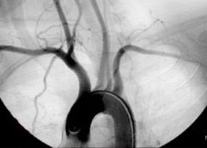
Aortography of the heart
This procedure is prescribed quite rarely. Despite the fact that this method is relatively easy to use, cardiac aortography by means of aortic puncture may be quite dangerous and cannot be offered for widespread use.
The essence of the problem with this procedure is that it makes no sense to conduct the examination using a small-diameter needle, while a medical instrument of a diameter suitable for the examination, due to its wide lumen, is not recommended due to the high probability of subsequent hemorrhage. This medical term means the outflow of blood from an injured vessel, a violation of its integrity and permeability of its walls. The presence of bleeding increases the risk of serious complications, or even the threat of death.
The choice of the injection site for this procedure - the brachial artery - does not help either. If the contrast fluid is injected through the above-mentioned blood vessel, the dye will have to travel a fairly long way before it reaches the required artery. This will not allow obtaining a picture of the required accuracy. But this method will protect the patient and the doctor from the risk of developing hemorrhage.
It is preferable to perform cardiac aortography through the carotid artery. The entire procedure is done fairly quickly, the substance is injected under high pressure to prevent the radiopaque agent from entering the brain. A series of images are taken at the moment of fluid injection.
This research method is quite innovative and is currently performed only in specialized institutions.
 [ 6 ], [ 7 ], [ 8 ], [ 9 ], [ 10 ], [ 11 ]
CT aortography
Computer tomographic angiography is, in fact, two in one. If the need arises and the patient is prescribed CT aortography, then the patient can undergo practically two examinations in one procedure: traditional scanning of the disturbing area using a computer tomography device, and angiography is performed in parallel. As a result, the specialist receives the most complete picture of pathological changes, and the series of images taken is copied and stored on the computer hard drive. The doctor will help, if necessary, to use this data repeatedly.
After the procedure, the attending physician will have in his hands high-precision images of the aorta, adjacent tissues and internal organs.
The procedure itself is similar to a regular CT scan. However, an additional feature is that during scanning, a contrast fluid is injected into a specific artery, after which several X-ray images are recorded.
Because the contrast agent is most often injected into a vein rather than an artery, CT aortography is considered less invasive than aortography alone.
The doctor may prescribe this examination in the same cases that were designated as indications for abdominal aortography. During the examination, the subject is placed on a special bed (the patient is on his back) and, using special mechanisms, is "brought" into a chamber - a cocoon. In it, the person's body is penetrated by a ring of X-rays.
Receiving a response, the computer program creates images - sections of various parts of the body. The resulting image is in black and white negative gradation. When the contrast agent enters the patient's body, the image becomes clearer. In this case, the doctor receives material in a three-dimensional (3D) image.
[ 12 ], [ 13 ], [ 14 ], [ 15 ], [ 16 ]
MRI aortography
Magnetic resonance angiography is a fairly innovative method of examining the human body. The combination of two methods - MRI aortography - allows a specialist to obtain an image of the organ of interest in one procedure, as well as X-ray images of this area.
The essence of the method is that the patient is placed in the magnetic field of the device, and his body is irradiated with radiological waves. The human body in such a situation responds with electromagnetic energy, which is recognized and processed by a computer program.
Magnetic resonance angiography is prescribed when there is a need to obtain a three-dimensional image of blood vessels. At the same time, this method allows you to obtain information and get a result without resorting to radiographic contrast agents. Although if there is a need for a clearer picture, doctors resort to using a contrast agent.
The advantage of this method is that it is painless. At the same time, doctors have not noted any negative impact of the magnetic field on the patient's body.
Seldinger aortography
One of the most frequently encountered and applied diagnostic methods, in the issue under consideration, is Seldinger aortography. This method of percutaneous catheterization of the femoral artery is performed using a set of special medical instruments. This set contains:
- A medical needle for performing a puncture.
- A metal conductor with a soft end.
- A dilator is an instrument for widening natural or artificially created openings and channels. It is especially relevant in the case of their reduction due to pathological changes in the patient's body.
- A catheter is a medical device consisting of a long thin tube and additional various attachments that allow it to perform various functions.
- An introducer is a “guide”, a plastic tube with a hemostatic valve built into it.
Before the examination, the patient undergoes a standard preparation procedure, which has already been described above. The examination itself begins with the insertion of a puncture needle into the femoral artery. This allows a special metal conductor (similar to a string) to be inserted into the puncture. The needle is removed, and using the "string", a medical catheter is fed into the artery's passage section.
To improve the clarity of the image, a contrast fluid is used, the quantitative introduction of which is calculated according to the formula 1 ml per kilogram of the patient's body weight (in some cases 2 ml per kilogram). As the monitoring of this study shows, such volumes do not provoke any complications and do not cause any harm to the patient's body.
Since this manipulation is quite painful, the patient is given anesthesia before it begins. Most often, the examination is carried out under the influence of a local anesthetic (lidocaine or novocaine), but under certain conditions and medical indications, general anesthesia can be used.
It is also worth noting that Seldinger aortography can be performed not only through the femoral aorta, but also through a puncture in the brachial or axillary artery. The doctor may decide to change the entry site due to blockage of the femoral blood vessel.
This technique is considered basic and is used most frequently in diagnosing the diseases listed above.
[ 17 ], [ 18 ], [ 19 ], [ 20 ], [ 21 ], [ 22 ], [ 23 ]
Translumbar aortography
If a doctor needs to visually examine the abdominal aorta or other large blood vessels that "serve" the pelvic organs and lower limbs of a person, then, often, he resorts to translumbar puncture. In case of blurring of the drawing and the need to obtain a clearer picture, doctors resort to the help of another examination procedure, called translumbar aortography.
Puncture of the blood vessel is performed with a special medical hollow needle. The introduction occurs from the back of the body. High-level translumbar aortography is also possible, in which case the catheter is inserted in the chest area at the level of the twelfth vertebra. If it is necessary to examine the work of the leg vessels (along their entire length) or the abdominal area, translumbar needle insertion occurs in the area of the second lumbar vertebra.
When carrying out the procedure in question, it is very important to comply with a number of mandatory requirements. One of these is the fact of the gradual removal of the needle:
- It is initially extracted directly from the aorta.
- And only after several minutes have passed can the instrument be removed from the para-aortic zone.
The staged removal helps prevent the formation of hemorrhages and hematomas in the para-aortic region.
This research method allows for a thorough examination of virtually any part of the arterial bed. The method is quite highly informative!
Research of this nature is necessarily carried out in specialized institutions. This makes it possible to reduce the risk of complications to a minimum, and the patient will receive assistance from highly qualified medical personnel.
Complications of abdominal aortography
Due to the fact that this examination is carried out using a medical instrument that damages the integrity of the skin and tissue structures of the body, and there is also damage to the blood vessel, there is a possibility of complications.
The most common complications of abdominal aortography are:
- Pain and swelling at the site of catheter insertion.
- The occurrence of bleeding. It can be of both internal and external nature.
- Formation of a hematoma.
- Blood vessel thrombosis.
- Arterial embolism (blockage of the vascular lumen by an embolus, i.e. a particle carried by the blood flow).
- Formation of an arterial or venous fistula.
- An allergic reaction to the iodine component of the contrast agent is possible.
- Development of aneurysm at the site of catheter insertion.
- Heart rhythm disturbances are possible.
- There is a risk of developing acute renal and/or hepatic failure.
- Perforation of a blood vessel.
Reviews of abdominal aortography
Thanks to the availability of the "world wide web", it is not difficult to obtain almost any information on a particular medical study. Forum discussions of the issue of interest are also becoming the norm.
Based on this, a person who is assigned to undergo the procedure in question may well familiarize themselves with its essence before undergoing it. It will not be difficult for any patient to read the article directly about the diagnostic examination itself. And also, reviews of abdominal aortography from those respondents who have already undergone this procedure will not be superfluous.
The reviews themselves are quite contradictory in terms of the procedure. Some complain of subsequent swelling and significant hematomas. But in general, a person does not experience significant negative sensations. There is no doubt that after undergoing this examination, the doctor receives a fairly informative picture of the pathological process occurring in the patient's body, which significantly reduces the time for establishing a diagnosis, and, accordingly, a faster start to treatment.
This diagnostic method has recently appeared among the "services" of doctors. Aortography is an innovative method of examining the aorta and other large blood vessels to identify a developing pathological process that negatively affects the patient's health. At the same time, the use of a radiopaque substance and appropriate equipment will allow you to get an instant series of clear contrast images that will help the specialist quickly make the correct diagnosis and begin treatment to stop the process as soon as possible.