
All iLive content is medically reviewed or fact checked to ensure as much factual accuracy as possible.
We have strict sourcing guidelines and only link to reputable media sites, academic research institutions and, whenever possible, medically peer reviewed studies. Note that the numbers in parentheses ([1], [2], etc.) are clickable links to these studies.
If you feel that any of our content is inaccurate, out-of-date, or otherwise questionable, please select it and press Ctrl + Enter.
Foot X-rays for adults and children
Medical expert of the article

Last reviewed: 03.07.2025
Almost everyone knows what radiography is. It is a specific and very common type of diagnostics that uses X-rays. However, not everyone knows in what cases this examination is prescribed - for example, when is it necessary to take an X-ray of the foot?
A foot X-ray can be prescribed by both a traumatologist and an orthopedist, depending on the problem with which the patient sought medical help. This type of diagnostics is indispensable for foot injuries and diseases: the study will help to clarify the diagnosis, determine the treatment tactics, and monitor the patient's recovery.
Indications for the procedure
According to statistics, the most common foot pathology is osteoarthritis: its "popularity" is associated with the mechanical production of cartilage (as a result of age-related changes or excess weight). But, in addition to osteoarthritis, foot X-rays are also prescribed for other inflammatory processes:
- rheumatoid arthritis;
- joint damage in psoriasis;
- ankylosing spondylitis;
- Reiter's disease.
Common foot pathologies also include gouty joint disease and diabetic neuropathic osteoarthropathy.
Tumor processes in the foot are relatively rare: as a rule, they are benign, and are most often represented by cysts or enchondromas. Radiography is the best way to diagnose such neoplasms.
X-rays of the foot are also prescribed for traumatic injuries, such as fractures of bone structures. Thus, X-rays allow us to determine the anatomy of the injury, its direction, linearity, and the degree of bone fragmentation; X-rays are also necessary to differentiate a fracture from a dislocation.
General indications for performing foot X-rays are:
- violation of bone integrity;
- tumor processes;
- flat feet;
- arthritis (rheumatoid, psoriatic, septic, osteoarthritis);
- Reiter's disease;
- osteoarthropathy, ankylosing spondylitis).
A common problem that requires radiographic confirmation is flat feet, in which the biomechanics of the foot is disrupted due to the loss of the ability to absorb shock. With flat feet, the ligamentous apparatus of the foot weakens, the arch becomes flatter, and the body weight is distributed from the heel area to the middle part of the foot. Due to pathologically altered biomechanics, the spine, ankle and hip joint become overload compensators. As a result, articular cartilage and intervertebral discs change, and joint deformation is observed. Externally, this is manifested by pain in the lumbar region, in the calf muscles, in the feet. In addition, complications may develop:
- arthrosis;
- varicose veins;
- heel spurs;
- curvature of the spine.
- An X-ray of the foot for flat feet may be prescribed if the patient voices the following complaints:
- pain in the foot, calves or back not associated with physical activity;
- external changes of the foot;
- frequent foot injuries;
- intense exercise, excess weight, hereditary predisposition to flat feet and foot deformities.
An X-ray of the foot is especially necessary for the military registration and enlistment office if the conscript has third-degree flat feet - it is with this degree of pathological changes that a person is considered unfit for military service. With the second degree of flat feet, the decision "fit with restrictions" may be announced.
 [ 1 ]
[ 1 ]
Preparation
Generally speaking, no specific preparation is required to perform a foot X-ray. Before going to the procedure, the patient should think about what clothes and shoes to wear so that the foot being examined can be quickly exposed in the office.
If a pregnant patient is referred for an X-ray, she must inform the doctor about her “condition”.
The entire foot X-ray procedure can last about fifteen minutes: the direct period of exposure to the rays does not exceed one second.
X-rays are taken in a special room – an X-ray room. An ambulatory patient can go for the examination on their own. Non-ambulatory patients and children may need help from relatives or parents. If someone accompanying them remains in the room during the procedure, they are asked to put on special protective clothing (an apron) to protect themselves from radiation.
The foot is placed on a table or a special stool until the required position is achieved. If several images are taken in different projections, the radiologist will periodically change the position of the patient's foot. In addition, an image of a healthy foot may be required (if a comparison is necessary).
Technique foot X-rays
During the X-ray of the foot, the patient does not feel anything - there are no pleasant or unpleasant sensations. The position of the foot that must be taken to obtain a correct image may seem not very comfortable: however, this is not a problem, because this position will only have to be held for a couple of seconds.
If the patient is unable to maintain the required position due to severe pain (for example, after an injury), the radiologist must help the patient determine another acceptable position that is more comfortable and no less informative.
After receiving the image, the X-ray doctor reviews the image, interprets it and sends the results to the attending physician.
The time it takes to obtain results may vary – from 1-2 hours to 1-2 days.
X-ray of the toes allows you to examine the structure and condition of the bone apparatus well, and analyze the quality of joint function. In what cases can the procedure be prescribed:
- if dislocations and fractures are suspected;
- for inflammatory diseases;
- in case of impaired blood circulation in the foot (especially in cases where the etiology cannot be determined);
- with impaired motor function of the fingers.
As a rule, X-rays of the toes are performed in two projections.
X-ray of the joints of the foot is often performed in relation to the entire ankle joint. The procedure is performed in different projections, depending on the diagnostic requirement and the patient's complaints, with or without the use of a load. The most informative in this situation are: lateral image of the foot, oblique image of the foot, image of the calcaneus.
X-rays of the foot joints can reveal:
- traumatic injuries;
- inflammatory diseases;
- degenerative processes;
- congenital bone and joint pathologies;
- osteophytes;
- metabolic and secondary disorders.
X-rays of both feet, left and right feet can be performed in cases of flat feet, as well as in cases where the doctor needs to compare both distal parts of the limbs. Depending on the patient's complaints and the suspected diagnosis, the doctor may require visualization of the feet in different positions:
- X-ray of the foot in lateral projection - this examination is performed in a lying or standing position, and the X-ray radiation is directed from the left angle (if the left limb is being examined) or from the right angle (when examining the right limb).
- Foot X-ray in two projections may include oblique and dorsal-plantar imaging. Oblique imaging is obtained when the patient places the foot on a special cassette with an inclination (the standard inclination angle is 45°, but it can be changed if necessary). Dorsal-plantar imaging is performed when the patient places the foot on a flat table, with a slight posterior deviation of the shin. In this case, the X-ray radiation should be directed from above.
- X-rays of the foot in a direct projection are often performed to diagnose flat feet, congenital or acquired deformities. Sometimes direct and anteroposterior projections are used to compare both feet, and they must be touching each other.
X-ray of the feet with load is usually prescribed if the patient complains of "unclear" pain in the limbs, without an obvious cause. This procedure is also in demand for flat feet, when the shape of the foot is disturbed. This type of examination is especially common in pediatric practice: it is used for early diagnosis of flat feet.
A load X-ray is performed in two projections. During the procedure, a person must stand on one leg, while bending the other at the knee, transferring the body weight to the limb being examined. The two projections include a direct and lateral image: the cassette is alternately positioned under the foot and on the side of the ankle joint. In most cases, both feet are examined.
To assess the functional capacity of the foot, the doctor may recommend taking images both with and without load: the position of the foot during such diagnostics should be the same.
X-ray of a child's foot
Children are prescribed foot X-rays no less frequently than adult patients: damage to the bone-ligament mechanism in childhood occurs mainly due to injuries, but the study is also used for congenital deformities, inflammatory processes, etc.
Many pathologies, including congenital ones, can often be eliminated if timely examination and treatment are carried out. For example, such serious problems as flat feet and clubfoot can be corrected in time.
For example, a child is said to have clubfoot when the child's foot is turned inward: there is typical plantar flexion. The emphasis falls on the outer surface of the foot, which is manifested by a change in gait.
Flat feet: such a diagnosis is given to a child only after the formation of the transverse and longitudinal arch of the foot is complete - that is, from about ten years of age. At an earlier age, the pathology can be corrected, so there is no need to make such a diagnosis.
X-ray of feet during pregnancy
Often, a pregnant woman faces the need to take an X-ray of her foot and doubts whether it will harm the future child. Indeed, such procedures are not welcomed during pregnancy, and are completely contraindicated during the first trimester. However, there is no need to worry: no one will take an X-ray for a woman without sufficient indications. And if such compelling indications do exist, the doctor will take all measures to protect the expectant mother and her child from the harmful effects of radiation.
The foot is relatively far from the abdominal area, so the impact of X-rays can be reduced to almost zero. For this, the woman will be asked to wear a special apron with a lead protective layer during the procedure. Upon arrival home, the patient should take a shower and drink a cup of milk. Usually, these measures are enough to neutralize the negative impact of the diagnosis on the body. Additionally, you can visit your gynecologist and consult with him: perhaps a repeat ultrasound will be recommended to assess the child's condition.
Contraindications to the procedure
The radiation produced during foot X-rays is considered completely safe for human health, provided that the procedures are infrequent. However, we must not forget about the conditions when X-ray examination is better replaced by other types of diagnostics.
Firstly, if possible, X-rays should not be performed on pregnant patients: they are performed only for vital emergency indications. Even if such a study is performed, the woman must first wear a special lead protective apron.
It is not advisable to have a foot X-ray if such a procedure has already been performed recently or several times. Frequent irradiation is extremely undesirable for the body. Therefore, you should not insist on the procedure if there are no appropriate indications for it.
There are no other contraindications to the study.
Normal performance
A high-quality X-ray of the foot allows you to examine this part of the limb in sufficient detail. Immediately after the procedure, the resulting image is carefully studied by a radiologist: his goal is not to make a diagnosis, but to describe what he saw with the recording of all detected pathologies. Then the image with the description is sent to the attending physician. It is he who makes the final diagnosis based on the results obtained, after which he determines the treatment tactics.
It is very important to carefully examine the image. For example, foot morphometry by X-ray is performed to diagnose longitudinal flatfoot: the doctor needs to make an additional measurement of the angle of the arch of the foot. The normal angle should not exceed 130°, with an arch height of at least 3.5 cm. When determining transverse flatfoot, a direct image of the foot is required. It is considered normal if only the heads of the I and V metatarsal bones are adjacent to the support.
If a clinic or diagnostic center has a modern X-ray machine, it will usually show all the nuances of the foot structure in more detail. This will allow the doctor to make an accurate diagnosis and prescribe the correct treatment.
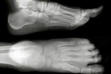
X-ray of a healthy foot, normal
The foot is a mobile mechanism represented by bones, joints, soft tissues. Thanks to this structure, a person has the ability to stand, walk, run or jump.
The skeleton of the foot is quite complex: it is completely “thought out” by nature to perform the function assigned to it.
During X-ray diagnostics, the device transmits radiation through the required area of the limb, and the resulting "picture" is transferred to a computer monitor or special X-ray film. The image displays all the bone elements and soft tissues that make up the structure of the foot: the ankle joint, the metatarsal system, and the finger phalanges.
As many people know, the X-ray "picture" is presented in white and black tones. At the same time, denser elements prevent the X-ray flow from passing through them - for example, bones, so they have a white color in the image. Soft structures (for example, muscle tissue) pass the rays through themselves and appear dark. Thus, the denser the structure, the lighter it is.
Typically, the radiologist performs the procedure in three projections: anterior-posterior, lateral and oblique image.
X-ray signs during foot examination
When describing certain pathological changes, the doctor uses various terms that characterize the current X-ray picture. However, there are no standard schemes for such descriptions: each radiologist has his own algorithms that he uses when making a conclusion. We can only name a number of signs by which the doctor determines traumatic, destructive and other processes in the bone-articular apparatus of the foot.
Thus, minor damages, such as bone cracks, may remain unnoticeable on X-rays. A more precise diagnosis can only be made after performing a CT scan.
A foot fracture has a specific appearance on an x-ray, and the typical signs are:
- line of enlightenment;
- fragmentary displacement;
- the arrangement of bone fragments at an angle.
In order to determine the treatment tactics, the doctor must assess the nature of the damage relative to the joint surface. An extra-articular fracture heals faster and is rarely accompanied by the development of complications. An intra-articular fracture affects the bones that are part of the joint structure. Such a violation often leads to a limitation of the motor ability of the foot; a bone callus may form. Such a callus has the appearance of an intense darkening focus.
Varus foot deformity may be present in several variants on X-ray. Flat-valgus deformity is typically characterized by pronounced changes in the bone structure of the middle and back sections, as well as the base of the metatarsal bones. If the pathology is congenital, the most pronounced are the disorders localized in the middle section. Deformed cuneiform, cuboid and navicular bones are detected against the background of moderate osteoporosis, large-loop images of bone trabeculae with rarefaction zones. Some trabeculae are thickened, with orientation along the load axis to the middle section of the foot. The calcaneus lacks the typical spongy structure. The IV and V bases of the metatarsal bones are especially deformed.
Equino-polovarus deformity is characterized by an increased longitudinal arch, calcaneal supination, absence of a transverse arch, hammer toes, equinus. The intensity of the bone pattern may decrease uniformly, and the bone trabeculae are thinned. Partial preservation of the lines of force in the talus and heel is observed. The head of the talus and the calcaneus form a large-loop image of trabeculae. Deformation of the navicular and cuneiform bones may be present, with the navicular bone displaced to the dorsal side. Osteoporosis is most pronounced in the heel bone (calcaneal tubercle).
Arthrosis of the feet on X-rays manifests itself differently, depending on many factors. In particular, early and chronic arthrosis are characterized by a number of special signs.
At the initial stage of development, arthrosis has the following symptoms:
- mild narrowing of the joint space;
- punctate calcifications;
- moderate signs of osteosclerosis.
In advanced arthrosis, the picture is somewhat different and expands:
- the joint space narrows significantly;
- the symptoms of osteosclerosis are more pronounced;
- bone tissue is compacted;
- subluxation is noticeable, the volume of the joint surface decreases, and flattening is observed;
- osteophytes are present
Arthritis of the feet on the X-ray is characterized by the widening of the joint space, which is explained by the presence of inflammatory effusion in the joint cavity. In addition, other signs are observed:
- compaction of soft tissues near the site of inflammation;
- deposition of calcifications.
Foot gout also looks like arthritis on an X-ray, but gout is also characterized by specific signs - for example, the presence of uric acid accumulation zones. The presence of urates is recorded in periarticular tissues, in the joint space: a clear structure of the joint surfaces is detected. In gouty arthritis, MRI diagnostics are more informative.
The diabetic foot on an X-ray is characterized by significant structural changes, pathological fractures, fragmentation and destruction of bone tissue (mainly the tarsal and metatarsal bones), divergence of joints, and secondary bone growths.
An ankle dislocation is another common injury, for the diagnosis of which in some cases it is necessary to resort to X-rays. With a dislocation, a change in the articular relationships of bone joints is observed. Dislocations and subluxations are distinguished - complete and incomplete displacements of joints. An X-ray of the foot quite clearly determines the nature and extent of pathological changes in the joint. It is possible to examine the condition of the periarticular tissues and bone damage. With traumatic dislocations, there are tears of the articular edges and bone sections, and all this should be visualized using X-rays. The study is performed in two projections. The most frequently diagnosed dislocations are the Lisfranc, Chopart joint, or isolated dislocations of individual bones.
Complications after the procedure
Foot X-rays are considered a safe diagnostic procedure. Despite the apparent health risks associated with X-rays, the amount of radiation used to take the image is not dangerous.
Radiologists use the minimum amount of radiation necessary to obtain the optimal diagnostic result.
Modern X-ray machines significantly surpass their predecessors in the quality of the resulting image and the dose of directed radiation. That is, the latest devices are much safer. The "picture" is displayed directly on the doctor's monitor, where he conducts an assessment without using additional radiation to the patient. It follows that for your own safety, it is advisable to have a foot X-ray performed in a good medical institution that has new, high-quality diagnostic equipment.
Doctors have not voiced the concept of the maximum acceptable dose of X-ray radiation for diagnostic purposes. Therefore, most often the procedure is prescribed exactly as many times as the doctor needs to make a diagnosis or to track the dynamics.
Of course, you can't be sure that foot X-rays will be safe if the diagnostics are carried out very often. But in many cases, X-rays become the only possible way to avoid major problems and complications that may be a consequence of one or another disease.
You should not ignore protection from X-rays. Today, three methods of such protection are known: time, distance and shielding. Thus, the duration of radiation exposure determines the amount of radiation dose received. The same can be said about the distance: the further the patient is, the smaller the dose he receives. A special screen installed between the patient and the X-ray machine also has a protective capacity. For this reason, it is advisable to use special "clothing" during diagnostics, such as lead aprons, caps, collars, etc.
Men and women who are planning to conceive a child are advised to protect their abdominal area and genitals from the rays.
When diagnosing children, it is generally advisable to cover the entire body, avoiding the area of the foot being examined.
Moreover, you should not perform more than one type of X-ray examination in one day (for example, you cannot perform a foot X-ray and a fluorography, or CT scan, or mammography, etc. on the same day).
Care after the procedure
After a single foot X-ray procedure, there is no point in taking any measures to care for and remove radiation from the body, as it is inappropriate. However, if a person has been exposed to X-rays several times in a row, then some post-procedural issues can be considered.
When you get home, you must take a shower.
There are a number of medications known to help the body cope with a small dose of radiation:
- Polyphepan – can be used in adult and pediatric practice;
- Potassium orotate – prevents the accumulation of radioactive cesium;
- dimethyl sulfide – has antioxidant properties;
- dietary supplements with calcium – accelerate the elimination of radioactive strontium.
In addition to taking medications, you need to focus on proper nutrition to speed up the body's cleansing from radiation.
Immediately after the foot X-ray procedure, you should drink a cup of milk - this product copes well with small doses of radiation. Dry wine or grape juice can be an alternative to milk.
Doctors advise drinking plenty of fluids, drinking fruit and vegetable juices, eating raw quail eggs, oatmeal, and dried fruits after the examination.
It is strongly discouraged to drink vodka to neutralize radiation. It has been proven that strong alcohol not only does not remove radioactive components, but also accelerates their distribution in the body tissues.
[ 12 ]
Foot X-ray Reviews
The X-ray examination method is considered the most frequently prescribed and accessible diagnostic method, which is used for various pathologies of the musculoskeletal system. X-ray can be classified as a relatively safe, easily tolerated method. In addition, it is also very informative: it helps to assess the condition of the bones, see the degree of injury or the nature of the pathological disorder.
X-rays of the foot are also indispensable for monitoring the dynamics of tissue healing after injuries and surgeries.
Generally speaking, an X-ray procedure often allows us to answer the question about the origin of pain in the legs and even in the back, to find out the reason for constant swelling of the legs and changes in the shape of the foot.
Foot X-ray is a procedure about which you can read only positive reviews. The method allows you to detect many pathologies hidden from the eye; it is accessible and always easily perceived by patients. Therefore, in traumatology and orthopedics, X-ray can be safely included in the first-priority series of procedures.