
All iLive content is medically reviewed or fact checked to ensure as much factual accuracy as possible.
We have strict sourcing guidelines and only link to reputable media sites, academic research institutions and, whenever possible, medically peer reviewed studies. Note that the numbers in parentheses ([1], [2], etc.) are clickable links to these studies.
If you feel that any of our content is inaccurate, out-of-date, or otherwise questionable, please select it and press Ctrl + Enter.
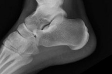
X-ray of the heel in two projections
Medical expert of the article

Last reviewed: 03.07.2025
The most accessible, informative and painless method of visualizing bone structures is radiography. The image also clearly shows damage to joints, cartilage of traumatic and inflammatory genesis, congenital defects. X-rays of the heels give an accurate idea of the presence or absence of damage after an injury, helps to determine the cause of discomfort in this area of the limb.
This procedure is used not only by trauma surgeons and orthopedists, but also by rheumatologists, endocrinologists to confirm connective tissue lesions, and oncologists – if a tumor in this location is suspected.
Indications for the procedure
- Suspected post-traumatic damage to bone, articular, and cartilaginous tissue in the heel area.
- The patient complains of discomfort in this localization, lameness, gait disturbances with suspected inflammatory processes (arthritis, bursitis, synovitis, osteomyelitis), degenerative changes (arthrosis, gout, heel spurs), the presence of congenital defects (flat feet, clubfoot, hallux valgus) or neoplasms of bone and joint tissue.
- For deep purulent abscesses on the back of the foot, to prevent the spread of infection to bone tissue.
- Monitoring treatment results.
Technique X-rays of the heels
An X-ray of the heel can be taken in different positions of the foot, the choice of which is made by the doctor based on the need to visualize it from a certain angle.
The axial projection of the calcaneus is performed most frequently. Typically, the patient lies on a table with straight legs, the film cassette is placed under the dorsum of the heel of the affected leg, and the foot is bent as much as possible toward the shin, sometimes with a bandage held by the patient himself. The central beam of X-rays is directed at the calcaneal tuberosity approximately at the median of the right angle with the surface of the table.
It is possible to take an axial projection image in a standing position. The patient places the foot of the affected leg on the film cassette, taking a position in which the shin is bent above the floor at an angle of approximately 45°, with the other leg placed back. The body position is fixed by supporting it on a nearby object, such as a chair. The X-ray beam is directed at the calcaneal tuberosity at an angle of 20° to the vertical axis.
To take a lateral projection image, the patient is placed on his side on the side of the limb being examined. A cassette is placed under his heel, the X-ray beam is directed vertically, and an image is taken. The other leg is bent slightly backwards, removing it from the X-ray zone.
There can be many options for laying down, depending on the capabilities of the equipment and the required shooting angle, for example, in a straight line - you can lie on your back, bend your knees and rest your feet on the table, or the patient lies on his stomach, with the heel of the sore leg up, and a bolster is placed underneath it at the level of the ankle joint.
X-ray diagnostics for arthritis to determine the degree of joint destruction is performed under load - the patient stands on the sore leg. If necessary, an X-ray of the heel of the second (healthy) leg is sometimes taken for comparison.
X-rays of the heel of a child are taken only when visualization is necessary, as, incidentally, with an adult. The technique is similar. The most difficult thing in this procedure is ensuring immobility. Small children are taken to the X-ray room with their parents, who calm the baby, hold them and ensure the necessary position and immobility of the limb. Vital areas of parents and children are protected with lead aprons during the procedure.
Contraindications to the procedure
For one X-ray procedure of the heels, the radiation dose on any equipment does not exceed 0.01 mSv. There are no absolute contraindications to X-ray diagnostics of the heel bone. Relative contraindications are pregnancy and childhood, when X-rays are done only in case of extreme necessity.
It is not recommended to perform the procedure on patients with severe bleeding and in critical conditions (shock, coma).
 [ 6 ]
[ 6 ]
Normal performance
An X-ray can show the internal structure of the heel bone, cartilage, joint connections, analyze the shape and relative position, and identify existing disorders – fractures, dislocations, degenerative and inflammatory changes.
X-rays of healthy heels show whole, even and dense parts of the calcaneus – the body and tubercle, with clear contours without roughness and patterns. X-rays of the heel normally do not contain darkening, displacement of the articular surfaces and proliferation of bone tissue (osteophytes, tumors), due to which the contour of the calcaneus becomes irregular. The cartilaginous pads have a normal thickness, the bones are not deformed.
X-ray: signs of disease
If you complain of heel pain, you must prescribe a radiological diagnosis. Pain may not be related to an injury, but it always indicates trouble. Radiography is the most widely available and informative method that gives an idea of the condition of bone tissue.
A fairly common cause of pain is plantar fasciitis or heel spur. The sharp wedge-shaped growth makes itself known by intense pain when a person steps on the heel, hyperemia and the appearance of a hard, light spot on the skin.
A heel spur is clearly visible on a lateral X-ray, since it is a bone formation. It looks like a wedge- or claw-shaped growth on the lower surface of the calcaneal tubercle, usually closer to its center. The growth is usually small in size, since with a spur more than 5 mm high, the patient can no longer walk due to severe pain. Although osteophytes of 20 mm are also common. An X-ray can often suggest the cause of the growth. Most often, it is flat feet; the appearance of a spur can also be caused by trauma or a tumor.
After an injury, an X-ray is prescribed to avoid missing a heel fracture. If it is detected, the location of the damage and the degree of complexity are determined.
A fresh injury is accompanied by severe pain and swelling in the heel area. X-rays are taken in two projections; fracture lines look like dark, uneven lines on white bones. The contours of the bones may remain (a fracture without displacement - a crack) or shift relative to each other. There is also a comminuted fracture, when the bone splits into several small pieces. All these types can usually be clearly seen on an X-ray.
There are cases when the fracture is not visible on the image, but the symptoms suggest its presence. Then, using the X-ray taken in the lateral projection, the Böhler angle is determined. Two straight lines are drawn. One of them is drawn through the upper points of the dorsal calcaneus and the subtalar joint. The other is drawn through the upper points of the subtalar joint and the frontal process of the calcaneus. The acute angle at the intersection of these lines is measured. If its value is less than 20°, then the presence of a fracture is assumed, for confirmation of which it is recommended to additionally do a CT scan.
The heel bone in a child can "break", held in place by the intact periosteum. Such a fracture has the shape of a twig.
Osteoporosis – rarefaction, decrease in bone density visually appears on the image as unevenness or roughness of the bone, change in its color, appearance of a pattern, since decalcified bones transmit X-rays, which illuminate the film.
Tumors of bones, cartilage tissue and mixed tumors appear on an X-ray as additional formations with unclear contours.
Complications after the procedure
If you follow the recommended rules, there will be no undesirable consequences after the procedure that may be associated with radiation exposure.
If there is a need to perform an X-ray of the heels of pregnant women, the abdomen is carefully covered with an apron with lead plates.
People in shock, pre-comatose, and comatose states are sensitive to even minimal doses, so even after injuries or accidents, X-rays are taken only when the patient’s condition has stabilized.
The same applies to patients with severe bleeding. A complication after the X-ray procedure may be a violation of blood flow, so diagnostics are not carried out until the condition stabilizes.
[ 9 ]
Reviews
Reviews of heel X-rays are positive. X-rays are quite informative, widely available, painless, and do not require special preparation. Compared to computed tomography, they are much cheaper, and the radiation dose during X-rays is ten times less. However, sometimes additional extensive diagnostics are required.