All iLive content is medically reviewed or fact checked to ensure as much factual accuracy as possible.
We have strict sourcing guidelines and only link to reputable media sites, academic research institutions and, whenever possible, medically peer reviewed studies. Note that the numbers in parentheses ([1], [2], etc.) are clickable links to these studies.
If you feel that any of our content is inaccurate, out-of-date, or otherwise questionable, please select it and press Ctrl + Enter.
Conjunctival examination
Medical expert of the article
Last reviewed: 06.07.2025

The conjunctiva is easily accessible for examination and diagnosis of many of its diseases and does not require any special equipment.
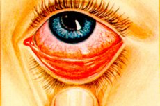
When examining the conjunctiva, it is necessary to pay attention to its color, transparency, shine, surface condition, presence of films, scars and discharge. Normal conjunctiva is pink, smooth, shiny and transparent (the meibomian glands are visible through it in the form of yellowish stripes, parallel to each other and perpendicular to the edge of the eyelid).
In case of inflammation of the conjunctiva ( conjunctivitis ), it acquires a rich bright red color and loses transparency due to the fact that its tissue swells (the meibomian glands are indistinguishable). The surface of the conjunctiva becomes rough and velvety due to the fact that the papillae, invisible to the naked eye in the normal conjunctiva, swell and enlarge; lymphatic follicles develop, which look like grayish-yellow nodules. Sometimes a film forms on the conjunctiva (in diphtheria and some acute conjunctivitis ). In some diseases ( trachoma, diphtheria, burns, etc.), scars appear on the conjunctiva - from minor superficial to coarse and extensive silvery-white scars. As a result of scarring, the conjunctiva shrinks and shortens, especially in the area of the transitional folds. The conjunctiva of the sclera also loses its luster and transparency during inflammation. On the eyeball, it is necessary to distinguish between superficial vessels and deep ones; thus, here one can observe the dilation of both superficial vessels - conjunctival injection, and deep vessels at the corneal limbus - pericorneal, or ciliary, injection. It is very important to distinguish between these two types of injections in diagnostic terms. Superficial, or conjunctival, injection indicates damage to the conjunctiva, while deep ciliary, or pericorneal, injection contributes to damage to the cornea and choroid.
In conjunctival injection, the conjunctiva is bright red; the dilated vessels move with the conjunctiva. Pericorneal injection is expressed mainly around the cornea; it refers to the deeper vessels lying in the superficial layers of the sclera; this hyperemia has a lilac or violet hue, and in this case the dilated vessels do not move with the conjunctiva.
If one or the other injection is present, we speak of a mixed injection.
It is necessary to pay attention to the presence of conjunctival discharge, which can be mucous, mucopurulent and purely purulent. If the amount of discharge is small, lumps are found on the conjunctiva, especially on the transitional folds, as well as at the corners of the eyes; with a large amount of discharge, the discharge flows over the edge of the eyelid, gets on the cheeks, sticks the eyelashes and eyelids together. If discharge is present, bacteriological studies are carried out to determine the nature of pathogenic microorganisms - a smear is examined or a culture is made on various nutrient media.
The clinical symptoms of common conjunctival diseases are so typical and the treatments so simple that recognition and treatment are not difficult for a non-specialist doctor. Under the supervision of a doctor, even mid-level health workers can treat conjunctival diseases.
 [ 1 ], [ 2 ], [ 3 ], [ 4 ], [ 5 ], [ 6 ], [ 7 ], [ 8 ], [ 9 ]
Laboratory studies of the conjunctiva
Indications
- Severe purulent conjunctivitis: identify infectious agents and initiate appropriate antimicrobial therapy based on the susceptibility of the infectious agent.
- Follicular conjunctivitis: differentiate viral from early chlamydial infection.
- Conjunctival inflammations, the clinical picture of which is not characteristic enough to accurately suggest etiological diseases.
- Conjunctivitis of newborns.
Special studies of the conjunctiva
- Tissue culture studies are now rarely performed, as they have been replaced by more accurate and rapid methods.
- Cytological examination, based on the detection of typical cellular infiltrates, is insensitive and subjective.
- Seeding of sensitive cell lines and observation of cytopathic effect or visualization with various chemicals and immunostaining methods.
- Detection of viral or chlamydial antigens in conjunctival and corneal preparations.
- Impression cytology: A cellulose acetate filter paper is pressed onto the conjunctiva or cornea, the surface epithelial cells adhere to the paper and are then examined. This helps in the diagnosis of ocular surface neoplasia, dry eye, ocular cicatricial pemphigus, limbal stem cell injury and infections.
- Polymerase chain reaction allows rapid identification of extremely small amounts of DNA with a very high degree of specificity. The reaction is used to detect adenovirus, herpes simplex virus and Chlamydia trachomatis in conjunctival smears.

