
All iLive content is medically reviewed or fact checked to ensure as much factual accuracy as possible.
We have strict sourcing guidelines and only link to reputable media sites, academic research institutions and, whenever possible, medically peer reviewed studies. Note that the numbers in parentheses ([1], [2], etc.) are clickable links to these studies.
If you feel that any of our content is inaccurate, out-of-date, or otherwise questionable, please select it and press Ctrl + Enter.
Corneal keratometry
Medical expert of the article

Last reviewed: 07.07.2025
Corneal keratometry is the measurement of the curvature of the axial meridians of the anterior surface of the cornea.
Optical principles of keratometry
- The cornea is a convex lens with a constant value of curvature for each meridian.
- Due to the properties of the cornea, the points projected by the device onto the surface of the cornea (two vertical and two horizontal) are reflected, which makes it possible to measure the radius of curvature (in mm) and convert it into diopters.
Limitations of Keratometry
- Keratometry makes it possible to measure the corneal surface limited to only four points located approximately 3 mm apart, but does not provide information about the central and peripheral zones of the cornea in relation to these points.
- Moderately expressed disturbances of the corneal surface can cause distortions and decrease the accuracy of measurements, therefore the method is practically not used for measuring non-spherocylindrical corneas, which are encountered in refractive surgery, keratoconus and other severe corneal pathologies.
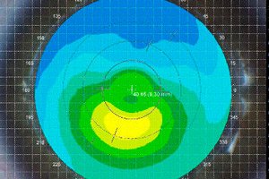
Study of corneal topography using computer videokeratoscopy allows obtaining a color coding map of the corneal surface and calculating the indices of the two main meridians (refractive power in diopters and their axes).
Indications for corneal topography examination
- Quantitative assessment of irregular astigmatism and corneal surface changes associated with contact lens wear.
- Early diagnosis of keratoconus, since diagnosis of its initial and preclinical manifestations is significantly difficult.
- Evaluation of postoperative corneal topography after refractive surgery, keratoplasty or cataract extraction.
Color scales
- Absolute scale: predetermined endpoint values; each scale color represents a specific diopter interval. Normal corneal topography maps are typically in the yellow-green spectrum. The values of this scale should be used to compare changes over time.
- The relative scale is not fixed and varies according to the diopter range of the particular cornea. It is important to study the scale before interpreting the chart.
 [ 1 ], [ 2 ], [ 3 ], [ 4 ], [ 5 ]
Evaluation of results
The interpretation of a keratotopogram is always based on practice. The following questions must be answered:
- What scale is used in keratotopogram?
- Is the scale appropriate?
- Is the keratotopogram reliable?
- What is the position of the pupil relative to the curvature pattern on the screen?
Who to contact?