All iLive content is medically reviewed or fact checked to ensure as much factual accuracy as possible.
We have strict sourcing guidelines and only link to reputable media sites, academic research institutions and, whenever possible, medically peer reviewed studies. Note that the numbers in parentheses ([1], [2], etc.) are clickable links to these studies.
If you feel that any of our content is inaccurate, out-of-date, or otherwise questionable, please select it and press Ctrl + Enter.
Additional methods of examination of the patient
Medical expert of the article
Last reviewed: 04.07.2025

By now, medicine has been enriched with a large number of additional research methods, the significance and distribution of which are gradually changing.
Laboratory methods. General blood tests and urine analysis retain their primary importance. Morphological examination of blood (primarily leukocytes) is of decisive importance in recognizing tumor processes - leukemia. Of no less importance is the quantitative determination of erythrocytes ( anemia ), leukocytes (severity of the inflammatory reaction), and measurement of the erythrocyte sedimentation rate ( ESR ).
Numerous studies of blood plasma and serum are carried out: biochemical, immunological, serological, etc. Some of them can be of decisive, key importance in diagnostics. These data reflect, in combination with other, primarily clinical manifestations, the course of pathological processes, a decrease or increase in their activity. It is possible to identify a complex of shifts indicating changes in protein fractions of the blood during active inflammatory and immune processes. An increase in the content of alanine and aspartic transaminases in the blood is observed in necrosis (death) of myocardial tissue ( infarction ), liver (hepatitis). Evaluation of the content of protein, glucose in urine, quantitative study of cellular elements in urine sediment have an important diagnostic value.
The study of feces, cerebrospinal fluid, and pleural fluids retains its importance in diagnostics. At the same time, it is especially necessary to stipulate the importance of bacteriological examination of all the listed environments, which often allows us to identify the etiological factor of the disease - the corresponding microorganism. Less important at present is the study of gastric juice and duodenal contents.
Instrumental methods. X-ray examination of various organs remains important in the diagnosis of diseases of the heart, lungs, gastrointestinal tract, gall bladder, kidneys, brain, and bones. Its accuracy and reliability of data have increased significantly with the use of so-called contrasting (barium suspension introduced into the gastrointestinal tract and contrast containing iodine introduced into the vascular bed).
The study of the electrical activity of some organs, primarily the heart (electrocardiography), is of great importance. It allows us to identify changes in the heart rhythm and pathology associated with morphological changes (hypertrophy of the heart, myocardial infarction ). Endoscopic examination has become especially important. Flexible endoscopes provide the opportunity to obtain good image quality and, thanks to a computer, allow us to carefully examine the inner surface of the gastrointestinal tract, bronchi, and urinary tract. An important, and sometimes decisive, addition to this study is a tissue biopsy with subsequent morphological study, which allows us to assess, for example, the malignancy of the process or the characteristics of the inflammation. Material for morphological examination can also be obtained by needle biopsy of the liver, kidneys, and myocardium.
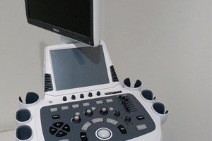
Ultrasound examination (echolocation) has become very popular in recent years. Ultrasound pulses, reflected from the boundaries of areas with different densities, allow obtaining information about the size and structure of organs. Ultrasound examination (ultrasound) of the heart is especially important, and it is possible to study its contractile function. Ultrasound of the abdominal organs, liver, gall bladder, and kidneys is also important. With the use of computers, the resolution of ultrasound and the quality of the images obtained have improved significantly. A very important advantage of ultrasound is its safety and non-invasiveness, which distinguishes it from angiography, liver biopsy, kidneys, and myocardium.
Computer tomography has made it possible to obtain high-quality images of dense organs and has acquired an important role in diagnostics. Radioisotope examination is used quite widely in the examination of the cardiovascular system, kidneys, liver, bones, and thyroid gland. A substance is introduced into the body that accumulates in the corresponding organ and contains a radioactive isotope, the radiation of which is subsequently recorded. In this case, morphological and functional deviations can be detected in the corresponding organ. Diagnostic studies are very diverse. Many of them are invasive, which raises the issue of examination safety. In any case, the danger of the studies conducted should not exceed the significance of the data that can be obtained.
Thus, in the diagnosis of human disease, the most important place still belongs to clinical examination, based primarily on classical methods. Although with the help of a number of additional and special research methods (laboratory, radiological and radiopaque, ultrasound, etc.) it is possible to clarify the features of changes in one or another organ, more accurately determine their localization (the location of stenosis of the coronary artery of the heart using coronary angiography, etc.) and even establish morphogenetic changes (various methods of studying tissue obtained during organ biopsy), the final diagnosis is still the result of a thorough comprehensive comparison of all the results obtained.


 [
[