If there is a need to assess the condition of the vascular network that feeds the brain area, ultrasound of the brachiocephalic arteries is prescribed.
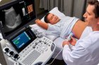
Transesophageal cardiac ultrasound, or transesophageal echocardiography, helps examine cardiac structures and assess cardiac function in more detail than standard ultrasound.
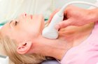
Among the many instrumental ultrasound diagnostic methods used by cardiologists, neurologists and surgeons, duplex scanning of the vessels of the head and neck is particularly common.
USDS, or ultrasound duplex scanning of the veins of the lower extremities, provides the physician with the ability to track baseline hemodynamic values.
Ultrasound is a diagnostic method that allows, using reflected ultrasound of certain frequencies, to visualize internal organs located, among other things, in the pelvic cavity: the bladder and rectum, the uterus with its appendages and the ovaries
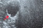
Any tissue area with increased density for ultrasound waves is a hyperechoic formation. Let's consider the causes of this phenomenon, types, diagnostic and treatment methods.
Ultrasound of the scrotum organs begins with the patient in a supine position using an ultrasound sensor with a frequency of at least 7 MHz. If it is necessary to visualize the dilated veins of the pampiniform plexus, the examination is also performed with the patient standing.