
All iLive content is medically reviewed or fact checked to ensure as much factual accuracy as possible.
We have strict sourcing guidelines and only link to reputable media sites, academic research institutions and, whenever possible, medically peer reviewed studies. Note that the numbers in parentheses ([1], [2], etc.) are clickable links to these studies.
If you feel that any of our content is inaccurate, out-of-date, or otherwise questionable, please select it and press Ctrl + Enter.
Heart
Medical expert of the article

Last reviewed: 06.07.2025
The heart (cor) is a hollow muscular organ that pumps blood into the arteries and receives venous blood. The heart is located in the thoracic cavity as part of the organs of the middle mediastinum. The heart is shaped like a cone. The longitudinal axis of the heart is directed obliquely - from right to left, from top to bottom and from back to front; two-thirds of it is located in the left half of the thoracic cavity. The apex of the heart (apex cordis) faces down, left and forward, and the wider base of the heart (basis cordis) faces up and back.
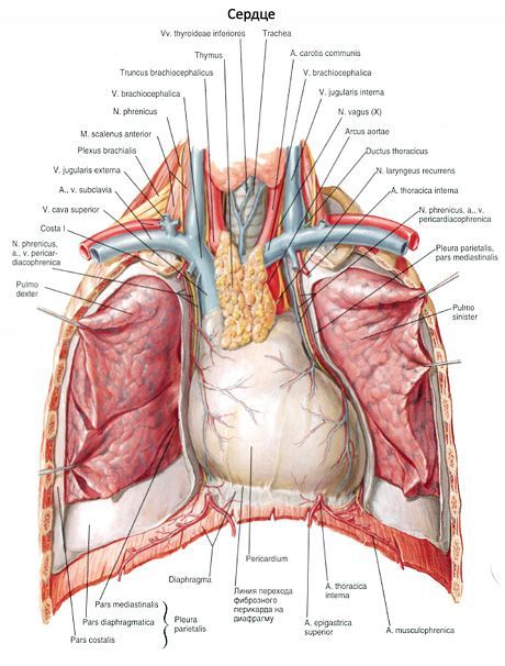
The sternocostal (anterior) surface of the heart (facies sternocostalis, s.anterior) is more convex, facing the back surface of the sternum and the cartilaginous parts of the ribs. The lower surface is adjacent to the diaphragm and is called the diaphragmatic surface (facies diaphragmatica, s.inferior). In clinical practice, this surface of the heart is usually called the posterior surface. The lateral surfaces of the heart face the lungs, each of them is called a pulmonary surface (facies pulmonalis). These surfaces (or edges) are visible in their entirety only when the lungs are moved away from the heart. On radiographs, these surfaces have the appearance of contours, the so-called edges of the heart: the right one is pointed and the left one is more obtuse. The average weight of the heart in men is approximately 300 g, in women - 250 g. The largest transverse size of the heart is 9-11 cm, the anteroposterior size is 6-8 cm. The length of the heart is 10-15 cm, the thickness of the wall of the atria is 2-3 mm, the right ventricle - 4-6 mm and the left - 9-11 mm.
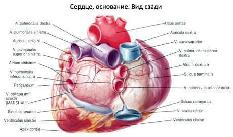
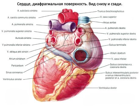
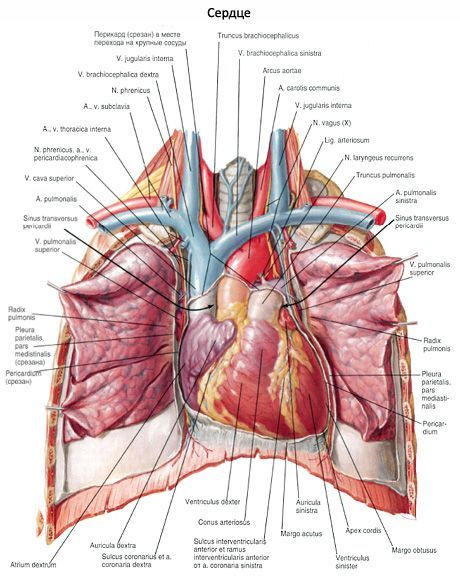
On the surface of the heart, there is a transversely located, rather deep coronary groove (sulcus coronarius), which is the border between the atria and ventricles. The coronary arteries of the heart are located in this groove. In front, the groove is covered by the pulmonary trunk and the ascending part of the aorta, behind which are the atria. Above the coronary groove on the anterior surface of the heart are part of the right atrium with its right auricle and the auricle of the left atrium, which lies entirely behind the pulmonary trunk. On the anterior sternocostal surface of the heart, the anterior interventricular groove (sulcus interventricularis anterior) is visible, to which the artery of the same name and the great cardiac vein are adjacent. On the back of the heart, the posterior interventricular groove (sulcus interventricularis posterior) is visible with the artery of the same name and the middle cardiac vein lying in it.
The longitudinal anterior interventricular groove divides the sternocostal surface of the heart into a larger right part, corresponding to the right ventricle, and a smaller left part, belonging to the left ventricle. The larger part of the left ventricle forms the posterior surface of the heart. The posterior (lower) interventricular groove begins on the posterior surface of the heart at the point where the coronary sinus enters the right atrium, reaches the apex of the heart, where it connects with the lower part of the anterior groove by means of the notch of the apex of the heart (incisura apicis cordis).
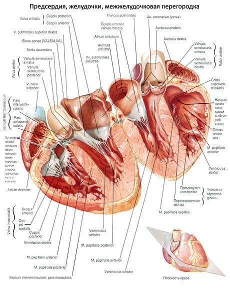
The heart consists of 4 chambers: two atria and two ventricles - right and left. The atria receive blood from the veins and push it into the ventricles. The ventricles eject blood into the arteries: the right one - through the pulmonary trunk into the pulmonary arteries, and the left one - into the aorta, from which numerous arteries branch off to the organs and walls of the body. The right half of the heart contains venous blood, the left half - arterial blood. The right and left halves of the heart do not communicate with each other. Each atrium connects to the corresponding ventricle through the atrioventricular orifice (right and left), each orifice is closed by cusp valves. The pulmonary trunk and aorta have semilunar valves at their beginning.
The right atrium (atrium dextrum), shaped like a cube, has a fairly large additional cavity - the right auricle (auricula dextra). It is separated from the left atrium by the interatrial septum (septum interatriale). On the septum, an oval depression is clearly visible - the oval fossa (fossa ovalis), covered by a thin membrane. This fossa, which is a remnant of the overgrown oval opening that connected the right and left atria in the fetus, is limited by the edge of the oval fossa (hmbus fossae ovalis). The right atrium has an opening of the superior vena cava (ostium venae cavae superioris) and an opening of the inferior vena cava (ostium venae cavae inferioris).
Along the lower edge of the opening of the inferior vena cava there is a small, inconstant, semilunar fold - the valve of the inferior vena cava (Eustachian valve; valvula venae cavae inferioris). In the embryo (fetus), this valve directs the blood flow from the right atrium to the left through the oval opening. Sometimes the valve of the inferior vena cava has a mesh structure: it consists of several tendinous threads connected to each other. Between the openings of the vena cava, a small intervenous tubercle (Lower's tubercle; tuberculum intervenosum) is visible, which is considered to be a remnant of the valve that directs the blood flow from the superior vena cava to the right atrioventricular opening in the embryo. The expanded posterior portion of the cavity of the right atrium, which receives both vena cavae, is called the sinus of the cavae (sinus venarum cavarum).
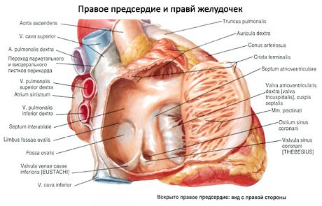
On the inner surface of the right auricle and the adjacent area of the anterior wall of the right atrium, longitudinal muscular ridges - pectinate muscles (mm.pectinati) protruding into the cavity of the atrium are visible. At the top, these ridges (muscles) end in a terminal crest (crista terminalis), which separates the venous sinus from the cavity of the right atrium (in the embryo, the border between the common atrium and the venous sinus of the heart passed here). The right atrium communicates with the ventricle through the right atrioventricular opening (ostium atrioventriculare dextrum). Between this opening and the opening of the inferior vena cava is the opening of the coronary sinus (ostium sinus coronarii). At its mouth, a thin crescent-shaped fold is visible - the valve of the coronary sinus (thebesian valve; valvula sinus coronarii). Near the opening of the coronary sinus there are pinpoint openings of the smallest veins (foramina venarum minimalum), which flow into the right atrium independently; their number may vary. There are no pectineal muscles around the opening of the coronary sinus.
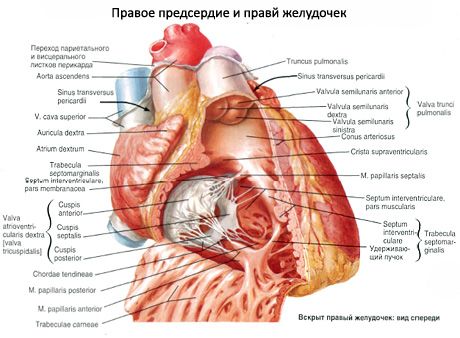
The right ventricle (ventnculus dexter) is located to the right and in front of the left ventricle, and is shaped like a triangular pyramid with the apex facing downwards. The slightly convex medial (left) wall of the right ventricle is formed by the interventricular septum (septum interventriculare), which separates the right ventricle from the left. The larger part of the septum is muscular (pars muscularis), and the smaller part, located in the uppermost section, closer to the atria, is membranous (pars membranacea).
The lower wall of the right ventricle, adjacent to the tendinous center of the diaphragm, is flattened, the anterior wall is convex anteriorly. In the upper, widest part of the ventricle there are two openings: behind - the right atrioventricular opening (ostium atrioventriculare dextrum), through which venous blood enters the ventricle from the right atrium, and in front - the opening of the pulmonary trunk (ostium trunci pulmonalis), through which blood is directed into the pulmonary trunk. The section of the ventricle from which the pulmonary trunk emerges is called the arterial conus (conus arteriosus). A small supraventricular ridge (crista supraventricularis) delimits the arterial conus from the inside from the rest of the right ventricle.
The right atrioventricular opening is closed by the right atrioventricular (tricuspid) valve (valva atrioventricularis dextra, s.valva tricuspidalis). The valve consists of three cusps: anterior, posterior and septal. The bases of the cusps are firmly fused with a dense connective tissue ring located on the border of the atrium and ventricle. The cusps of the atrioventricular valve are triangular folds of the inner lining of the heart (endocardium), into which fibrous fibers from the connective tissue ring extend. The free edges of the cusps, similar in appearance to thin tendon plates, face the cavity of the ventricle. The anterior valve cusp (cuspis anterior) is fixed on the anterior semicircle of the opening, the posterior valve (cuspis posterior) on the posterolateral semicircle, and finally, the smallest of them, the medial septal cusp (cuspis septalis), is fixed on the medial semicircle. When the atria contract, the cusps are pressed against the walls by the blood flow and do not interfere with its passage into the ventricular cavity. When the ventricles contract, the free edges of the cusps close, but do not evert into the atrium, since they are held in place on the ventricular side by dense connective tissue strands that stretch - the chordae tendineae.
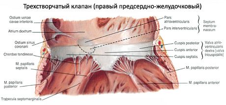
The inner surface of the right ventricle (except for the arterial cone) is uneven, here you can see the strands protruding into the lumen of the ventricle - fleshy trabeculae (trabeculae cdrneae) and cone-shaped papillary muscles (mm.papillares). From the top of each of these muscles - anterior (the largest) and posterior (mm.papillares anterior et posterior) - the majority (10-12) of the tendinous chords begin. Sometimes some of the chords originate from the fleshy trabeculae of the interventricular septum (the so-called septal papillary muscles). These chords are attached simultaneously to the free edges of two adjacent cusps, as well as to their surfaces facing the ventricular cavity. Therefore, when the atrioventricular valve closes, the cusps are set at the same level. Sometimes the chords are attached to the surfaces of the cusps facing the ventricular cavity.
Immediately at the beginning of the pulmonary trunk, on its walls, is the pulmonary trunk valve (valva trunci pulmonalis), consisting of three semilunar flaps located in a circle: anterior, left and right (valvulae semilunaris anterior, dextra et sinistra). The convex (lower) surface of the flaps faces the cavity of the right ventricle, and the concave (upper) and free edge - into the lumen of the pulmonary trunk. The middle of the free edge of each of these flaps is thickened due to the so-called nodule of the semilunar valve (nodulus valvulae semilunaris). The nodules contribute to a tighter closure of the semilunar flaps when they close. Between the wall of the pulmonary trunk and each of the semilunar flaps there is a small pocket - the lunula (sinus) of the semilunar valve (lunula valvulae semilunaris). During contraction of the ventricular muscles, the semilunar valves (valves) are pressed by the blood flow to the wall of the pulmonary trunk and do not impede the passage of blood from the ventricle. When the muscles relax, when the pressure in the ventricular cavity drops, the return flow of blood fills the lunulae (sinuses) and opens the valves: the edges of the valves close and do not allow blood to pass into the cavity of the right ventricle.
The left atrium (atrium sinistrum), which has an irregular cuboid shape, is separated from the right atrium by a smooth interatrial septum. The oval fossa located on the septum is more clearly expressed on the side of the right atrium. The left atrium has 5 openings, four of which are located above and behind - these are the openings of the pulmonary veins (ostia venarum pulmonalium), two on each side. The pulmonary veins do not have valves. The fifth opening is the largest; this left atrioventricular opening communicates the left atrium with the ventricle of the same name. The anterior wall of the left atrium has an anteriorly facing cone-shaped expansion - the left auricle (auricula sinistra). The inner wall of the left atrium is smooth, since the pectineal muscles are located only in the atrial auricle.
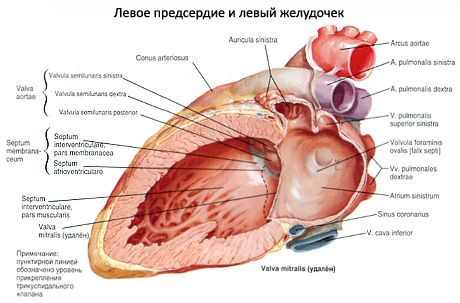
The left ventricle (ventriculus sinister) is cone-shaped, with the base facing upward. In the upper, widest section of the ventricle there are two openings. Behind and to the left is the left atrioventricular opening (ostium atrioventriculare sinistrum), and to the right of it is the aortic opening (ostium aortae). In the left atrioventricular opening there is the left atrioventricular valve (mitral valve; valva atrioventricularis sinistra, s.valva mitralis).
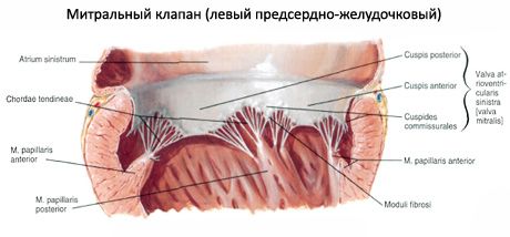
This valve consists of two triangular shaped cusps: the anterior cusp (cuspis anterior), which begins at the medial semicircle of the opening (near the interventricular septum), and the posterior cusp (cuspis posterior), smaller than the anterior one, beginning at the lateral-posterior semicircle of the opening.
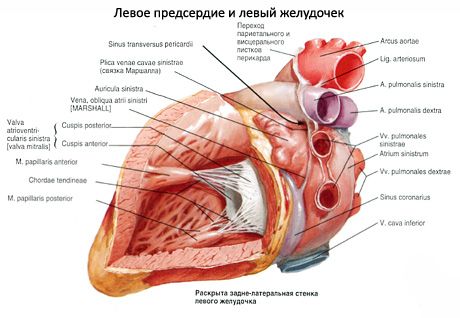
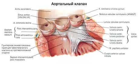
On the inner surface of the left ventricle (especially in the region of the apex of the heart) there are many large fleshy trabeculae and two papillary muscles - anterior and posterior (mm.papillares anterior et posterior). These muscles are located on the corresponding walls of the ventricle. Thick tendinous chords extend from the tops of the muscles, attaching to the cusps of the atrioventricular valve. Before entering the aortic opening, the surface of the ventricle is smooth. The aortic valve (valva aortae), located at its very beginning, consists of three semilunar valves: posterior (valvula semilunaris posterior), right (valvula semilunaris dextra) and left (valvula semilunaris sinistra). Between each valve and the wall of the aorta there is a small hole (sinus) of the semilunar valve (lunula valvulae semilunaris). The aortic valves also have nodules - nodules of the semilunar valves, located in the middle of the free edges; the nodules of the aortic valves are larger than those of the pulmonary trunk.
 [
[ Where does it hurt?
What's bothering you?
What do need to examine?