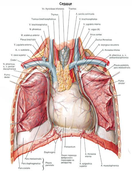
Pericardium
Pericardium (pericardium, pericardium) delimits the heart from neighboring organs, is a thin and dense, strong fibrous-serous sac, in which the heart is located. In the pericardium, two layers of different structures are distinguished: the outer layer is fibrous and the inner one is serous. The outer layer - the fibrous pericardium (pericardium fibrosum) near the large vessels of the heart (at its base) passes into their adventitia. Serous pericardium (pericardium serosum) has two plate - parientalnuyu (lamina parietalis), which lines the inside of the fibrous pericardium, and visceral (lamina visceralis, s.epicardium), which covers the heart, as its outer shell - epicardium. The parietal and visceral plates pass into each other in the region of the base of the heart, in the place where the fibrous pericardium is fused with the adventitia of large vessels: the aorta, the pulmonary trunk, and the hollow veins. Between the parietal plate serous pericardium outside and visceral lamina (epicardium) has a slit-like space - pericardial cavity (cavitas pericardialis), covering the heart from all sides and contains a small amount of liquid, the liquid wets the surface of the serous pericardium and provides them slipping during contraction of the heart. Serous pericardium is a thin plate, formed by a dense fibrous connective tissue, rich in elastic fibers. From the pericardial cavity, the serous pericardium is lined with flat epithelial cells - mesothelium; these cells are located on the basement membrane. Fibrous pericardium is formed by a dense fibrous connective tissue with a high content of collagen fibers.
Pericardium in shape resembles an irregular cone, the base of which (the lower part) is tightly fused with the tendon center of the diaphragm, and at the top (at the apex of the cone) embraces the initial sections of the large vessels: the ascending part of the aorta, the pulmonary trunk, and the upper and lower hollow and pulmonary veins. The pericardium is divided into three sections. The anterior sternum-costal section is connected to the posterior surface of the anterior thoracic wall by the sternocarpal ligament (ligamenta sternopericardiaca). It occupies the area between the right and left mediastinal pleurae. The lower part is diaphragmatic, fused with the sinewy center of the diaphragm. The mediastinal department (right and left) is the most significant in length. From the lateral sides and from the front, the mediastinal section of the pericardium is closely fused with the mediastinum pleura. Left and right between the pericardium and pleura pass the diaphragmatic nerve and the pericardial diaphragm vessels adjacent to it. Behind the mediastinal section of the pericardium is attached to the esophagus, the thoracic part of the aorta, the unpaired and semi-uneven veins, surrounded by a loose connective tissue.
In the pericardial cavity between him, the surface of the heart and large vessels are quite deep pockets - sinuses. First of all, it is the transverse sinus of the pericardium (sinus transversus pericardii) located at the base of the heart. Front and top, it is limited to the initial section of the ascending aorta and the pulmonary trunk, and from behind - the anterior surface of the right atrium and the superior vena cava. The oblique sinus of the pericardium (sinus obliquus pericardii) is located on the diaphragmatic surface of the heart. It is limited by the base of the left pulmonary veins on the left and the inferior vena cava on the right. The anterior wall of this sinus is formed by the posterior surface of the left atrium, the posterior by the pericardium.
In a newborn, the pericardium is spherical (rounded), and the heart is tight. The volume of the pericardial cavity is insignificant. The upper border of the pericardium is very high, along the line connecting the sternoclavicular joints; the lower border corresponds to the lower border of the heart. The pericardium of the newborn is mobile, since the sternum-pericardial ligaments that fix the adult pericardium are poorly developed. By the age of 14, the pericardial boundary and its relationship with the mediastinum organs are similar to those of an adult.
Vessels and nerves of the pericardium
In the blood supply of the pericardium, the pericardial branches of the thoracic part of the aorta, the branches of the pericardial diaphragmatic artery and the branches of the upper diaphragmatic arteries participate. Pericardial veins adjacent to the same-named arteries flow into the brachiocephalic, unpaired and semi- unpaired veins. Lymphatic vessels of the pericardium are directed to the lateral pericardial, prepericardial, anterior and posterior mediastinal lymph nodes. Pericardial nerves are the branches of the diaphragmatic and vagus nerves, as well as the cervical and thoracic cardiac nerves that extend from the corresponding nodes of the right and left sympathetic trunks.
Last reviewed: 25.06.2018