Ultrasound diagnosis (ultrasound)

prostate ultrasound

An ultrasound of the prostate (prostate gland) allows you to get an idea of its size, shape, structure, as well as the features of its relationship with other pelvic organs.

Ultrasound of the ankle joint

It should be noted that with the advent of new broadband and high-frequency sensors, the information content of ultrasound examination of tendons and ligaments of the ankle joint has increased significantly and the ultrasound method (ultrasound) today has an advantage over MRI.

Knee ultrasound

Today, the diagnostic capabilities of ultrasound examination (US) of the knee joint are being revised. Despite the fact that ultrasound methods in the study of the knee joint cannot completely replace such highly informative methods as MRI and standard X-ray examination, nevertheless, the ultrasound method has its own certain advantages.

Hip ultrasound in adults

Ultrasound of the hip joints can be an additional method to clinical or X-ray examinations. It should be noted that ultrasound is more informative than MRI in detecting small effusions in the hip joint, even less than 1 ml.

Ultrasound of the wrist and hand joints

The ultrasound method (US) has a number of advantages over magnetic resonance imaging in examining the soft tissues of the wrist and hand joints. Several points can be highlighted that highlight these advantages. Firstly, it is the convenience of ultrasound and the ability to quickly compare symmetrical sections.

Elbow ultrasound

Since the elbow joint is relatively small and superficially located, it is very convenient for examination using the ultrasound method (US). It can even be said that ultrasound is the method of choice for examining this joint due to its ease of implementation, information content and cost-effectiveness.

Shoulder ultrasound

In most medical institutions, X-ray examination of the shoulder joint remains mandatory in the algorithm for examining patients with shoulder pathology. It is well known that X-ray examination is highly informative when searching for traumatic injuries to bone structures.

Bone ultrasound

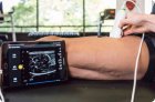
It is impossible to study the bone structure using the ultrasound method. However, the ultrasound method can be used to evaluate the bone surface and cortex. Targeted examination of the bone surface is carried out in rheumatoid arthritis, trauma, and various infections. Marginal erosions and synovial ulcers are best detected by ultrasound examination.

Joint ultrasound

A joint is a complex organ consisting of a capsule, synovial membrane, cartilage and bone. In addition, there are ligaments, tendons and muscles to stabilize the joint. Not all joints are the same in structure.

Nerve ultrasound

The emergence of new high-frequency matrix and wide-band sensors, new technologies for processing ultrasound signals (tissue harmonics, compound scanning) has provided ultrasound with priority in the study of peripheral nerves. It is customary to correlate the course of a nerve with its projection onto the skin.