
All iLive content is medically reviewed or fact checked to ensure as much factual accuracy as possible.
We have strict sourcing guidelines and only link to reputable media sites, academic research institutions and, whenever possible, medically peer reviewed studies. Note that the numbers in parentheses ([1], [2], etc.) are clickable links to these studies.
If you feel that any of our content is inaccurate, out-of-date, or otherwise questionable, please select it and press Ctrl + Enter.
Skull
Medical expert of the article

Last reviewed: 04.07.2025

The skull (cranium) is the skeleton of the head. It is the most complexly structured part of the skeleton, which serves as a receptacle for the brain, organs of vision, hearing and balance, smell and taste, and as a support for the initial sections of the digestive and respiratory systems. The human skull is formed by 23 bones (8 paired and 7 unpaired).
The skull is divided into the cerebral section, or cranial skull, and the facial, or visceral skull. The cerebral section of the skull (cerebral skull) is located above the facial section and contains the brain. The cranial skull (cranium cerebrale) is formed by the frontal, occipital, sphenoid, parietal, temporal and ethmoid bones and their joints. The facial section of the skull - the facial skull (cranium viscer&le) is represented by the bones of the masticatory apparatus: the upper and lower jaws, as well as small bones of the skull, which are part of the walls of the eye sockets, nasal and oral cavities. A special place is occupied by the hyoid bone, located in the anterior region of the neck.
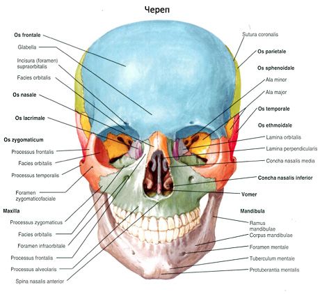
Bones of the cranial region of the skull
The frontal bone (os frontale) is involved in the formation of the anterior part of the vault (roof) of the skull, the anterior cranial fossa and the eye sockets. The frontal bone is divided into the frontal squama, orbital and nasal parts.
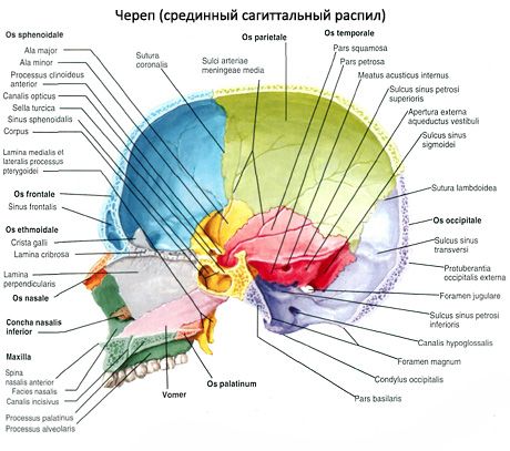
The sphenoid bone (os sphenoidale) occupies a central position in the base of the skull. It participates in the formation of the base of the skull, its lateral sections and a number of cavities and pits. The sphenoid bone is composed of the body, pterygoid processes, greater and lesser wings.
The occipital bone (os occipitale) is located in the posterior lower part of the cranial section of the skull. This bone is divided into the basilar part, two lateral parts and the occipital squama, which surround the large (occipital) opening (foramen magnum).
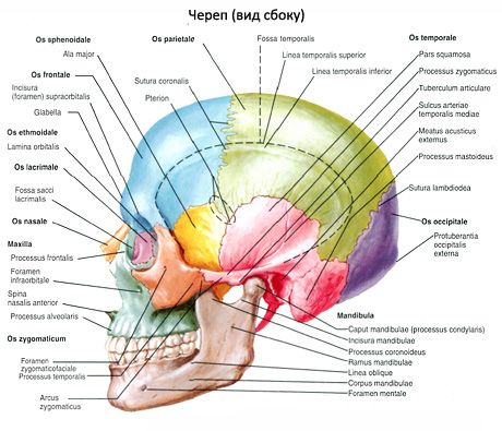
The parietal bone (os parietale) is paired, wide, convex outward, and forms the upper-lateral sections of the cranial vault. The parietal bone has 4 edges: frontal, occipital, sagittal, and squamosal. The frontal edge borders the posterior surface of the frontal squama, the occipital edge - with the occipital squama. The two parietal bones are connected to each other by the sagittal edge. The lower, squamosal edge is obliquely cut and covered by the squama of the temporal bone. The parietal bone has 4 angles: the anterior-superior frontal angle, the posterosuperior occipital angle, the anterior-inferior sphenoid angle, and the posteroinferior mammillary angle.
The temporal bone (os temporale) is paired and forms part of the base and lateral wall of the skull between the sphenoid bone in front and the occipital bone behind. It contains the organs of hearing and balance. The temporal bone is divided into the pyramid, tympanic and squamous parts.
 [ 1 ]
[ 1 ]
Bones of the facial skull
The upper jaw (maxilla) is a paired bone. The upper jaw has a body and four processes: frontal, alveolar, palatine and zygomatic.
The palatine bone (os palatinum) is paired and participates in the formation of the hard palate, orbit, and pterygopalatine fossa. It has two plates - horizontal and vertical, connected almost at a right angle, and three processes.
The inferior nasal concha (concha nasalis inferior) is a paired, thin curved plate with a body and three processes. The lateral surface of the body is fused with the upper edge of the concha crest of the upper jaw and the perpendicular plate of the palatine bone. All the processes of this concha extend from its upper edge.
The vomer is an unpaired bone plate that participates in the formation of the bony nasal septum. The lower edge of the vomer fuses with the nasal crests of the maxilla and palatine bone. The posterior edge of the vomer separates the choanae from each other. The anterior edge of the vomer is connected at the top with the perpendicular plate of the ethmoid bone, and at the bottom - with the cartilaginous nasal septum.
The nasal bone (os nasale) is paired and participates in the formation of the bony bridge of the nose. The upper edge of the nasal bone is connected to the nasal part of the frontal bone, the lateral edge - to the frontal process of the maxilla. The nasal bone also participates in the formation of the piriform aperture - the anterior opening of the nasal cavity.
The lacrimal bone (os lacrimale) is paired and forms the anterior part of the medial wall of the orbit. Below and in front, it is connected to the frontal process of the maxilla, and behind - to the orbital plate of the ethmoid bone. Above, the lacrimal bone borders on the medial edge of the orbital part of the frontal bone. On the lateral surface of the bone is the posterior lacrimal crest (crista lacrimalis posterior). Anterior to the lacrimal crest is the lacrimal groove (sulcus lacrimalis), which, together with the groove of the same name in the maxilla, forms the fossa of the lacrimal sac (fossa lacrimalis).
The zygomatic bone (os zygomaticum) is paired and connects the frontal, temporal and maxillary bones, strengthening the facial skull. The zygomatic bone has lateral, temporal and orbital surfaces. The lateral surface faces forward and laterally, contains a small zygomaticofacial opening (foramen zygomaticofaciale). The temporal surface forms the anterior wall of the infratemporal fossa, has a small zygomaticotemporal opening (foramen zygomaticotemporale). On the orbital surface, which forms the lower lateral wall of the orbit, there is also a small zygomaticoorbital opening (foramen zygomaucoorbitale).
The lower jaw (mandibula) is the only movable bone of the skull. The unpaired lower jaw has a body and two branches.
The hyoid bone (os hyoideum) is located in the anterior region of the neck, between the lower jaw at the top and the larynx at the bottom. It consists of an arched body and two pairs of processes - small and large horns. The short small horns extend upward, backward and laterally to the right and left of the body of the bone. The longer large horns, thickened at the ends, extend backward and slightly upward from the body of the bone. The hyoid bone is suspended from the skull by muscles and ligaments and is connected to the larynx.
Head movements occur at the atlanto-occipital joint around the frontal, sagittal and vertical axes.
Extension of the head (tilting the head back) is performed by: the trapezius, sternocleidomastoid, splenius, semispinalis and longissimus capitis muscles, the large and small posterior rectus capitis muscles, and the superior oblique capitis muscles.
Flexion of the head (forward tilt) is performed by: the long muscles of the head, the anterior rectus muscles of the head, the lateral rectus muscles of the head, as well as the suprahyoid and infrahyoid muscles (with a fixed lower jaw).
Tilt of the head to the side (right, left) occurs with the simultaneous contraction of the extensor muscles and flexor muscles of the corresponding side.
Rotational movements (turns) of the head together with the atlas to the right or left (in the medial and lateral atlantoaxial joints) around the odontoid axial vertebra are performed by the following muscles: the splenius capitis, the longissimus capitis, the inferior oblique capitis on its side, and the sternocleidomastoid muscle on the opposite side.
Muscles that move the lower jaw in the temporomandibular joints. Raise the jaw: temporal muscles, masseter muscles, medial pterygoid muscles. Depress the lower jaw: digastric muscles, geniohyoid muscles, mylohyoid muscles, infrahyoid muscles. Forward movement of the lower jaw: digastric muscles, geniohyoid muscles. Backward movement of the lower jaw (protruding forward): temporal muscles (posterior bundles). Sideways movement of the lower jaw: lateral pterygoid muscle (of the opposite side).
[ 2 ]
Использованная литература