
All iLive content is medically reviewed or fact checked to ensure as much factual accuracy as possible.
We have strict sourcing guidelines and only link to reputable media sites, academic research institutions and, whenever possible, medically peer reviewed studies. Note that the numbers in parentheses ([1], [2], etc.) are clickable links to these studies.
If you feel that any of our content is inaccurate, out-of-date, or otherwise questionable, please select it and press Ctrl + Enter.
The palatine bone
Medical expert of the article

Last reviewed: 04.07.2025
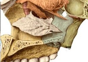
The palatine bone (os palatinum) is paired and participates in the formation of the hard palate, orbit, and pterygopalatine fossa. It has two plates - horizontal and vertical, connected almost at a right angle, and three processes.
The horizontal plate (lamina honsontalis) is fused with the same edge of the same plate of the palatine bone on the opposite side by its medial edge. The posterior edge of the horizontal plate is free, and the soft palate is attached to it. The anterior edge of the plate is connected to the posterior edge of the palatine process of the maxilla. As a result, the palatine processes and horizontal plates of the palatine bones form a hard bony palate (palatum osseum) on the entire skull.
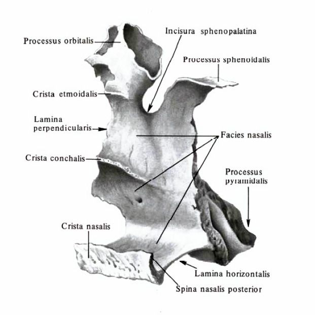
The perpendicular plate (lamina perpendicularis) participates in the formation of the lateral wall of the nasal cavity. On the lateral surface of this plate is the large palatine groove (sulcus palatinus major). Together with the grooves of the same name of the maxilla and the pterygoid process of the sphenoid bone, it forms the large palatine canal (canalis palatinus major). On the medial surface of the perpendicular plate there are two horizontal ridges. The upper ethmoid ridge (crista ethmoidalis) serves to attach the middle nasal concha, and the lower concha ridge (crista conchalis) - the inferior nasal concha.
The palatine bone has orbital, sphenoid and pyramidal processes.
The orbital process (processus orbitalis) is directed forward and laterally and participates in the formation of the lower wall of the orbit.
The sphenoid process (processus sphenoidalis) is oriented posteriorly and medially. It connects with the lower surface of the body of the sphenoid bone. The orbital and sphenoid processes delimit the sphenopalatine notch (incisura sphenopalatine), which, together with the body of the sphenoid bone, delimits the sphenopalatine foramen.
The pyramidal process (processus pyramidalis) extends from the palatine bone downwards, laterally and backwards. Narrow minor palatine canals (canales palatini minores) pass through this process, opening through openings on the palatine surface of the pyramidal process.
The maxillary, or maxillary, sinus (sinus maxillaris) is a cavity of the upper jaw. The anterior wall of the sinus is thin in the center, thickening in the peripheral sections. This wall is formed by the part of the upper jaw between the infraorbital margin and the alveolar process. The posterolateral wall corresponds to the tubercle of the upper jaw. The nasolacrimal canal is adjacent to the anterior section of the medial wall of the maxillary sinus, and the ethmoid cells are adjacent to the posterior section. The lower wall of the sinus is formed by the alveolar process of the upper jaw. The upper wall of the sinus is also the lower wall of the orbit. The maxillary sinus opens into the middle nasal passage. The sinus varies in shape and size.
The frontal sinus (sinus frontalis) varies considerably in size. The septum dividing the frontal sinus into right and left parts is usually asymmetrical. The frontal sinus communicates with the middle nasal passage.
The sphenoid sinus (sinus sphenoidalis) is located in the body of the sphenoid bone. The lower wall of the sinus participates in the formation of the wall of the nasal cavity. The cavernous sinus is adjacent to the upper part of the lateral wall. The sphenoid sinus is usually divided into two asymmetric parts by the sagittal septum. Sometimes the septum is absent. The sphenoid sinus communicates with the superior nasal passage.
The air cavities communicating with the nasal cavity are the anterior, middle and posterior cells of the ethmoid bone.
The bony palate (palatum osseum) is formed by the palatine processes of the right and left upper jaws, connected along the midline, and the horizontal plates of the palatine bones. It serves as a hard (bony) base for the upper wall of the oral cavity. The bony palate is limited in front and on the sides by the alveolar processes of the upper jaws, which form the upper alveolar arch. The median palatine suture (sutura palatina mediana) runs along the midline of the bony palate. At the anterior end of the palate is the incisive canal (canalis incisivus) for the nerve of the same name. Along the junction of the posterior edge of the palatine processes of the upper jaws with the horizontal plates of the palatine bones is the transverse palatine suture (sutura palatina transversa). In the lateral sections of this suture, at the base of each horizontal plate, there is an opening of the large palatine canal and 2-3 small palatine openings, through which the oral cavity communicates with the pterygopalatine fossa.
The upper and lower alveolar arches together with the teeth, as well as the body and branches of the lower jaw, form the skeleton of the anterior and lateral walls of the oral cavity.
Behind the maxilla is the infratemporal fossa (fossa infratemporalis), which is delimited from the temporal fossa at the top by the infratemporal crest of the greater wing of the sphenoid bone. The upper wall of the infratemporal fossa is made up of the temporal bone and the greater wing of the sphenoid bone (infratemporal crest). The medial wall is formed by the lateral plate of the pterygoid process of the sphenoid bone. The anterior wall of this fossa is the tubercle of the maxilla and the zygomatic bone. From the lateral side, the infratemporal fossa is partially covered by the branch of the mandible. In front, the infratemporal fossa communicates with the orbit through the inferior orbital fissure, and medially through the pterygomaxillary fissure (flssшra pterygomaxillaris) - with the pterygopalatine fossa.
The pterygopalatine fossa (fossa pterygopalatina) has 4 walls: anterior, superior, posterior and medial. The anterior wall of the fossa is the tubercle of the maxilla, the superior wall is the lower lateral surface of the body and the base of the greater wing of the sphenoid bone, the posterior wall is the base of the pterygoid process of the sphenoid bone, and the medial wall is the perpendicular plate of the palatine bone. From the lateral side, the pterygopalatine fossa communicates with the infratemporal fossa. Below, the pterygopalatine fossa gradually narrows and passes into the greater palatine canal (canalis palatinus major), which is limited below by the maxilla (laterally) and the palatine bone (medially). Five openings open into the pterygopalatine fossa. Medially, this fossa communicates with the nasal cavity through the sphenopalatine foramen, above and behind - with the middle cranial fossa through the round opening, behind - with the area of the lacerated opening by means of the pterygoid canal, and downwards - with the oral cavity through the greater palatine canal.
The pterygopalatine fossa communicates with the orbit via the inferior orbital fissure.
The bony palate (palatum osseum) is formed by the palatine processes of the right and left upper jaws, connected along the midline, and the horizontal plates of the palatine bones. It serves as a hard (bony) base for the upper wall of the oral cavity. The bony palate is limited in front and on the sides by the alveolar processes of the upper jaws, which form the upper alveolar arch. The median palatine suture (sutura palatina mediana) runs along the midline of the bony palate. At the anterior end of the palate is the incisive canal (canalis incisivus) for the nerve of the same name. Along the junction of the posterior edge of the palatine processes of the upper jaws with the horizontal plates of the palatine bones is the transverse palatine suture (sutura palatina transversa). In the lateral sections of this suture, at the base of each horizontal plate, there is an opening of the large palatine canal and 2-3 small palatine openings, through which the oral cavity communicates with the pterygopalatine fossa.
The upper and lower alveolar arches together with the teeth, as well as the body and branches of the lower jaw, form the skeleton of the anterior and lateral walls of the oral cavity.
What do need to examine?
How to examine?