
All iLive content is medically reviewed or fact checked to ensure as much factual accuracy as possible.
We have strict sourcing guidelines and only link to reputable media sites, academic research institutions and, whenever possible, medically peer reviewed studies. Note that the numbers in parentheses ([1], [2], etc.) are clickable links to these studies.
If you feel that any of our content is inaccurate, out-of-date, or otherwise questionable, please select it and press Ctrl + Enter.
Cuneiform bone
Medical expert of the article

Last reviewed: 06.07.2025
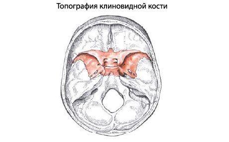
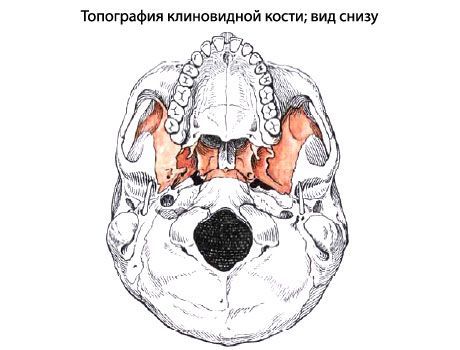
Sphenoid bone(os sphenoidale) occupies a central position in the base of the skull. It participates in the formation of the base of the skull, its lateral sections and a number of cavities and pits. The sphenoid bone is composed of the body, pterygoid processes, greater and lesser wings.
The body of the sphenoid bone (corpus sphenoidale) has an irregular shape and six surfaces: the upper, lower, posterior, fused (in an adult) with the basilar part of the occipital bone, anterior and two lateral surfaces. On the upper surface of the body there is a depression - the sella turcica (sella turcica) with a deep pituitary fossa (fossa hypophysialis). At the back of the sella turcica there is the back of the sella (dorsum sellae), and at the front - the tubercle of the sella (tuberculum sellae). On each side of the body of the bone there is a carotid groove (sulcus caroticus) - a trace of adjacency of the internal carotid artery. On the anterior surface of the body of the sphenoid bone there is a wedge-shaped ridge (crista sphenoidalis). On the sides of the ridge there are irregularly shaped wedge-shaped conchae (conchae sphenoidales), limiting the apertures of the sphenoid sinus. The sphenoid sinus (sinus sphenoidalis) is an air-filled cavity that communicates with the nasal cavity.
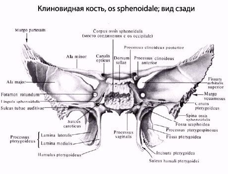
The lateral surfaces of the body of the sphenoid bone directly pass into the paired small and large wings.
The lesser wing (ala minor) is a laterally flattened bone plate, at the base of which is the optic canal (canalis opticus), leading into the eye socket. The posterior free edge serves as the border between the anterior and posterior cranial fossae. The anterior edge connects with the orbital part of the frontal bone and the cribriform plate of the ethmoid bone. Between the lesser wing at the top and the upper edge of the greater wing is an elongated opening - the superior orbital fissure (fissura orbitalis superior), connecting the cranial cavity with the eye socket.
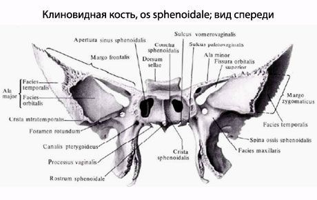
The greater wing (ala major) begins from the lateral surface of the body of the sphenoid bone with a wide base and, like the lesser wing, is directed laterally. It has four surfaces: cerebral, orbital, temporal and maxillary. The concave cerebral surface faces the cranial cavity. It has three openings through which blood vessels and nerves pass. The round opening (foramen rotundum), located closer to the base of the greater wing, leads into the pterygopalatine fossa. At the level of the middle of the wing is the oval opening (foramen ovale), opening on the base of the skull, and behind it is a small spinous opening (foramen spinosum). The orbital surface (facies orbitalis) is smooth and participates in the formation of the lateral wall of the orbit. On the temporal surface (facies temporalis) there is an infratemporal crest (crista infratemporalis), oriented in the anteroposterior direction and separating the temporal fossa from the infratemporal on the lateral surface of the skull.
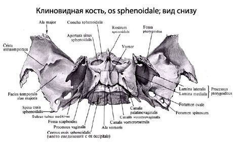
The maxillary surface (facies maxillaris) faces forward - into the pterygopalatine fossa.
The pterygoid process (processus pterygoideus) is paired and extends downwards from the body of the sphenoid bone. The process consists of medial and lateral plates (lamina medialis et lamina lateralis). Behind, between the plates, is the pterygoid fossa (fossa pterygoidea). At the base of the pterygoid process, from back to front, is the narrow pterygoid (vidian) canal (canalis pterygoideus), connecting the pterygopalatine fossa with the area of the foramen lacerum on the whole skull.
Occipital bone(os occipitale) is located in the posterior lower part of the cranial part of the skull. This bone is divided into the basilar part, two lateral parts and the occipital squama, which surround the large (occipital) opening (foramen magnum).
The basilar part (pars basilaris) is located in front of the large (occipital) opening. In front, it connects with the body of the sphenoid bone, together with which it forms a platform - a slope (clivus). On the lower surface of the basilar part there is an elevation - the pharyngeal tubercle (tuberculum pharyngeum), and along the lateral edge there is a groove of the inferior petrosal sinus (sulcus sinus petrosi inferioris).
The lateral part (pars lateralis) is paired and passes into the squama of the occipital bone at the back. Below each lateral part there is an elliptical elevation - the occipital condyle (condylus occipitalis), at the base of which is the hypoglossal nerve canal (canalis nervi hypoglossi). Behind the condyle there is a condylar fossa (fossa condylaris), and at its bottom is the opening of the condylar canal (canalis condylaris). To the side of the occipital condyle is the jugular notch (incisura jugularis), which, together with the jugular notch of the pyramid of the temporal bone, forms the jugular foramen. Next to the jugular notch on the cerebral surface is the groove of the sigmoid sinus (sulcus sinus sigmoidei).
The occipital squama (squama occipitalis) is a wide, outwardly convex plate with strongly serrated edges. On the whole skull, they are connected to the parietal and temporal bones. In the center of the outer surface of the squama, the external occipital protuberance (protuberantia occipitalis externa) is visible, from which a weakly expressed superior occipital line (linea nuchae superior) extends in both directions. The external occipital crest (crista occipitalis externa) runs down from the protuberance to the large (occipital) opening. From its middle, the inferior occipital line (hinea nuchae inferior) runs to the right and left. The highest occipital line (linea nuchae suprema) is sometimes visible above the external occipital protuberance.
On the inner side of the occipital squama is a cruciform eminence (eminentia cruciformis), dividing the medullary surface of the squama into 4 pits. The center of the cruciform eminence forms the internal occipital protuberance (protuberantia occipitalis interna). To the right and left of this protuberance runs the groove of the transverse sinus (sulcus sinus transversus). Up from the protuberance runs the groove of the superior sagittal sinus (sulcus sinus sagittalis superioris), and down, to the large (occipital) opening, runs the internal occipital crest (crista occipitalis interna).
 [
[ What do need to examine?
How to examine?