
All iLive content is medically reviewed or fact checked to ensure as much factual accuracy as possible.
We have strict sourcing guidelines and only link to reputable media sites, academic research institutions and, whenever possible, medically peer reviewed studies. Note that the numbers in parentheses ([1], [2], etc.) are clickable links to these studies.
If you feel that any of our content is inaccurate, out-of-date, or otherwise questionable, please select it and press Ctrl + Enter.
Pigmentless melanoma of the skin: symptoms, confusion, prognosis
Medical expert of the article

Last reviewed: 04.07.2025
Melanoma or skin cancer is one of the most common and most dangerous types of cancer. This disease tends to metastasize, and metastases appear very quickly, unlike other types of cancer, with which people live for several years. And the mortality rates from it are simply off the charts. And the worst thing is that this disease primarily affects young people aged 25-45. The key to successful treatment in the case of melanoma is its early diagnosis. But how can we nip the disease in the bud if we have a non-pigmented melanoma, not always noticeable even to an experienced eye?
Epidemiology
Among all patients diagnosed with malignant neoplasms, patients with melanoma are considered the rarest category, because skin cancer occurs 10 times less frequently than other cancers.
Achromatic melanoma is the rarest type of skin cancer. And this is good news, because this type of oncology is considered the most dangerous due to the rapid spread of metastases throughout the body. By the way, according to statistics, achromatic melanoma develops more often in women than in men or children.
This type of melanoma is also dangerous because in 20 percent of cases the disease is detected at late stages, when the metastasis process takes on generalized forms. The survival rate of patients with non-pigmented melanoma is slightly higher than 50 percent, while pigmented melanoma can be cured in more than 70 percent.
 [
[ Causes pigmentless melanoma
As we already know, melanoma tends to appear on the site of moles or in their vicinity. This makes it easy to notice it by changes in the appearance of the mole: its color, contour, skin characteristics. It is not for nothing that dermatologists recommend examining the marks we inherited daily, noting any changes in them, and if there are a large number of moles, undergoing an examination and dermatoscopy annually.
The situation is a little different with amelanotic melanoma, which, although a rarer phenomenon, is no less dangerous due to its unpleasant feature of metastasizing even at the initial stages of the disease. But if in the case of ordinary pigmented melanoma we are talking about the degeneration of cells of a mole, which is initially prone to this, then what causes pathological changes in ordinary skin cells in the case of amelanotic melanoma?
Risk factors
Doctors cannot yet answer this question precisely, because “black holes” can be found even in studies on pigmented (achromatic) melanoma. It is practically impossible to say specifically what caused the degeneration of a more or less pigmented area of the skin. We can only talk about the risk factors for the development of this pathology.
Such factors, as in the case of ordinary melanoma, include:
- Skin type. Melanoma is more often diagnosed in people with fair skin, blue eyes, light hair, and very often freckles.
- UV radiation. This includes both excessive exposure to the sun and regular visits to solariums. It turns out that lovers of a beautiful, rich tan are more susceptible to skin cancer than those who are satisfied with their natural skin tone.
- Increased solar activity. Being outdoors at this time and the sun's rays hitting exposed areas of the body are a presumed cause of the development of amelanotic or pigmented melanoma.
- Sunburn. We are not talking about severe thermal burns with blisters filled with liquid, but about severe skin irritation, accompanied by redness, itching, peeling, and in some cases the appearance of blisters, the sloughing off of the upper layers of the skin. Almost everyone is familiar with this phenomenon, especially at the beginning of the beach season, when many, not having calculated the intensity of the sun's rays, run to the pharmacy for "Panthenol" or to the store for sour cream. But melanoma can be a consequence not only of fresh burns, but even those received in childhood.
- Scars and trophic ulcers on the skin. Such formations are more susceptible to the appearance of malignant neoplasms than normal skin cells.
- Congenital sensitivity to ultraviolet rays. A rare inherited disorder called xeroderma pigmentosum is characterized by the appearance of large areas of intensely pigmented, deep brown tissue on the skin, which is considered more prone to degeneration than skin with normal pigment.
- Intraepidermal carcinoma or Bowen's disease.
- Paget's cancer, which appears as an inflamed, red spot.
- Borderline nevi (moles with dark borders, irregular shape, blurred edges, raised above the skin surface, etc.). Amelanotic melanoma can develop near such moles.
- Disruptions in the endocrine system. High levels of sex hormones, and estrogen in particular, can trigger pathological changes in cells and their uncontrolled growth.
- Large build. Tall, overweight people have a large area of skin, and the larger the area of skin, the more likely it is that some part of it will undergo degeneration. It is not for nothing that melanoma is diagnosed extremely rarely in children.
- Young and mature age.
- Weak immunity.
- Pregnancy and lactation periods. During this time, the skin and the entire body become more sensitive to the effects of irritants, which include UV radiation.
- High radiation background. Radiation is known to be one of the main causes of various mutations, including intracellular ones.
- Regular exposure of the skin to electromagnetic fields or chemicals. People who, due to their occupation, consistently experience such negative effects are more likely to get melanoma than others.
- Hereditary predisposition. The probability of malignancy (malignant transformation) of cells is higher in those people who have cases of oncology in their family (and not only skin cancer).
Usually, several factors are involved in the development of achromatic melanoma. Thus, a person with light skin and eyes, who avoids prolonged contact with sunlight, may never know what melanoma is, while his dark-skinned friend, who has a passion for a beautiful tan and a small scar on his skin, may suddenly end up on the surgeon's table with melanoma.
As for the causes of melanoma development, we cannot help but dwell on such a point as preventive removal of moles that have a risk of degenerating into melanoma. Many readers mistakenly think that after the above procedure, the risk of developing melanoma in the same place increases. In fact, this is not true. On the contrary, removing the risk factor only increases the chances of a healthy life.
Amelanotic melanoma after mole removal may appear near the surgical site or far from it, or not appear at all. The development of skin cancer in this case will not be related to the operation to remove the nevus.
It is a different matter if a small section of the border nevus was not removed during the operation. It is believed that the risk of cell degeneration at the site of removal becomes higher, but there have been no cases of malignancy in connection with the removal of moles.
We are talking about removing moles with clean histology, i.e. not containing cancer cells. Otherwise, one hundred percent guarantees cannot be given.
Pathogenesis
As in the case of pigmented melanoma, the pathogenesis of the achromatic form of skin cancer is not well understood. In essence, the elevation on the skin is a cluster of melanocytes that are unable to produce melanin in sufficient quantities. It is believed that cell mutations occurring at the gene-molecular level are to blame. Under the influence of some of the factors listed above, the structure of DNA and its enzyme system change, deviations from the norm are observed in chromosomes, the number of genes and their characteristics change.
By the way, scientists have long noticed that malignancy of cells can also be promoted by improper nutrition and the use of certain medications (hormonal drugs, oral contraceptives). As for food, the development of skin cancer is promoted by a diet with an excess of animal fats and proteins (mainly meat). An identical situation is observed with a deficiency in the diet of plant products, which are sources of vitamin A and C.
Symptoms pigmentless melanoma
The first signs of achromatic melanoma are considered to be: the appearance of a small elevation (tubercle) on the skin and its rather rapid growth. Externally, the tubercle resembles a small wart, so it may not attract attention for a long time. Moreover, its color is not much different from the color of the skin.
Achromatic melanomas can be flesh-colored, slightly pinkish or whitish. When the neoplasm begins to grow, roughness and exfoliated scales of the epithelial layer of the skin appear on its surface, the skin becomes rougher. In some cases, it looks like a scar, since it does not have smooth edges. When pressed, the seal does not cause discomfort or pain, so patients soon forget about the incomprehensible discovery on the body.
In appearance and shape, a pigment-free melanoma may resemble a small, almost flat elevation, resembling a colorless mole, or it may have a dome-shaped structure (nodular or nodular a pigment-free melanoma). In the second case, cell growth occurs vertically, i.e. the melanoma does not grow in width, but in height. Conventional flat melanomas are small in size, while nodular melanoma can be more voluminous and large.
A distinctive feature of non-pigmented melanoma is the uneven growth of its parts, due to which a noticeable asymmetry of the compaction is observed from the outside. The edges of a flat neoplasm are usually uneven, and the pigmentation is uneven.
An exception is the nodular form of achromatic melanoma, which is a dome-shaped, light, slightly rough or warty neoplasm that has a symmetrical shape and uniform color.
As the melanoma grows, it begins to cause discomfort. It may begin to hurt and itch. Redness and swelling of the tissues are observed at the site of the nodule and around it. The surface of the melanoma often begins to crack and bleed, and small ulcers form on it. This indicates the progression of the disease and its transition to later stages, which are very difficult to treat.
If there were hairs on the lump initially, and then they began to fall out, this is also an alarming symptom, indicating malignancy of the process. Very often, an increase in lymph nodes near the neoplasm is also noted.
Stages
The type of melanoma and the patient's condition depend entirely on the stage of development of the malignant process. It is customary to distinguish 4 stages of skin cancer:
- The first or initial stage of melanoma is characterized by the appearance on the skin of a convex, painless lump no more than 2 mm thick, located in the upper layers of the epidermis.
- The second stage is when the neoplasm thickens, penetrates deeper into the upper layers of the dermis, but metastases are not detected.
- At the third stage, the skin on the surface of the tubercle begins to peel, crack and bleed, and an increase in lymph nodes is noted, which indicates the penetration of cancer cells into the lymphatic system.
- The last fourth stage of melanoma is the spread of metastases to various organs and systems of the body. What does non-pigmented melanoma look like at the last stage? This is a seal with an uneven warty surface, sometimes reaching 3 or more centimeters, covered with a flaky crust with pinpoint hemorrhages and ulcers. Around it there is a hyperemic area of healthy skin. The seal causes constant discomfort: it hurts and itches. In color, it can remain pinkish or, in the case of a nodular form, acquire an almost black shade.
Most often, non-pigmented melanoma is diagnosed at stage 3 or even 4 of the disease, because before that it may not show itself in any way, having a resemblance to a harmless wart. Survival at stage 4 of the disease is almost zero. But even getting rid of melanoma at an earlier stage does not guarantee that after a while the disease will not return, but in a different place.
Forms
As we have already understood, achromatic melanoma can have different forms, and its symptoms depend on the stage of the disease. Let's consider the 2 most common types of achromatic melanoma:
- Superficially spreading type. The most common form of melanoma, characteristic of both pigmented neoplasms and those that have arisen on the skin without intense pigmentation. It is diagnosed in approximately 3 out of 4 cases of melanoma.
This melanoma looks like a plaque with uneven color and structure, uneven edges and relatively slow growth. It can be in the superficial layers of the skin for about 4-5 years, and then go deeper, metastasizing to various organs.
In men, such melanomas appear mainly on the arms, in women – on the legs.
- Nodular type. It is much less common, but is considered the most aggressive due to its tendency to grow quickly. It often appears in places where there are no moles. The papule-shaped neoplasm may remain light as the disease progresses, but more often it becomes dark in color. It quickly increases in size (in a few months), begins to ulcerate and bleed.
The nodular form of melanoma can also be located on the face, neck, back and other parts of the body. By the way, this form of the disease occurs slightly more often in men than in women.
A subtype of nodular melanoma is its desmoplastic variant, which looks like a hard, irregularly shaped node, strongly resembling a scar or cicatrice. It is very difficult to diagnose this form of melanoma, because even a superficial biopsy can be inclined towards a keloid or dermatofibroma, which have nothing in common with oncology.
Complications and consequences
Amelanotic melanoma is one of the most insidious oncological pathologies. Disguised as other skin diseases, it parasitizes in the body of an unsuspecting person until it completely disables the patient's body, affecting various organs and systems, reducing immunity and leaving the person no chance.
The absence of obvious symptoms of the disease at the early stages leads to the fact that melanoma progresses calmly, grows and spreads its pathologically altered cells throughout the body, where they again divide uncontrollably and disrupt the functioning of various organs. Metastases of non-pigmented melanoma, depending on its location, can be found in the lymphatic system, brain, cervical region, lungs, bones and other human organs and systems.
Cancer cells are carried throughout the body by the lymphatic system. Enlarged lymph nodes against the background of unusual compaction indicate the beginning of the metastasis process. If a tumor without metastases can be removed completely, then treating cancer with metastases is very difficult, because cancer cells can appear in different organs at any time.
Radiation and chemotherapy are not very effective in this case. In most cases, they allow the process to be slowed down a little, but there is no talk of a complete cure. The patient dies sooner or later.
Diagnostics pigmentless melanoma
It is not so easy to diagnose skin cancer, and it does not matter whether melanoma develops against the background of a pigmented nevus or on the surface of unchanged tissues. In the first case, at the initial stage of the disease, it resembles a common mole, in the second - a wart or a lipoma. It is for this reason that, having discovered areas of altered tissue, people are in no hurry to run to the doctor and do not associate their appearance with cancer.
No matter how much they say on the Internet about the dangers of melanoma, in real life the disease is very rare. This relaxes people even more, who believe that if the probability of getting sick is low, then their evil fate will certainly pass them by.
The chances of recognizing melanoma right away are quite slim. For example, a therapist who sees a small flesh-colored bump on a patient's skin that doesn't bother the person in any way is unlikely to attribute its appearance to the onset of cancer. Even a dermatologist may well confuse a colorless lump with a wart if the patient doesn't emphasize that the bump has begun to grow.
With suspicious seals on the skin that tend to increase, it is most logical to immediately contact a dermato-oncologist. The alarming symptoms (FIGARO rule) in this case are:
- Form. A flat neoplasm rises above the surface of the skin, i.e. changes shape.
- Change in size and other parameters. The tumor grows and changes its appearance.
- Borders. If the seal initially had smooth outlines, then over time the borders become blurred, the edges acquire an irregular and unclear shape.
- Asymmetry. If you mentally divide a bulge on the body in half, one half will noticeably differ from the other in thickness and shape.
- Size. The tumor not only grows, it becomes quite large.
- Coloring. The following should be alarming: uneven color of the neoplasm, the appearance of inclusions of other colors, the formation of a red or dark rim.
When you contact a doctor with your problem, you must tell him when the strange lump appeared, how it behaved, when and how actively it began to grow, whether there were cases of cancer in the family, etc. The doctor will definitely prescribe blood and urine tests, which will provide information about the patient's body condition and the presence of tumor markers (protein molecules, waste products of cancer cells) in the blood. If there are ulcers on the surface of the neoplasm, a smear from the damaged surface will be taken for cytological analysis.
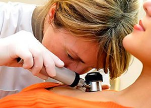
In the case of non-pigmented melanoma, it is very difficult to determine the nature of the neoplasm with the naked eye. Instrumental diagnostics of the disease begin with examining the compaction using a dermatoscope with the output of an enlarged image of the suspected melanoma on the computer screen. Sometimes an epiluminescent microscope is used for these purposes, which allows one to see the state of the neoplasm under the epidermis.
If enlarged lymph nodes are observed, scintigraphy or radioisotope examination, and in some cases, surgical biopsy of the lymph node, can help to identify its connection with the penetration and division of cancer cells.
A biopsy of the tumor tissue could provide more information, but it is not always possible. For example, in the case of an aggressive nodular form of skin cancer, a biopsy cannot be performed before surgery, since it can cause rapid growth of an already rapidly growing tumor. Very often, a biopsy, and then a histological analysis of the tissue taken from the affected area, is performed after surgery to remove the tumor. The material for the study is taken immediately during the surgery.
If the diagnosis confirms the presence of cancer cells in the neoplasm, it becomes necessary to check various organs for metastases. For this purpose, the following may be prescribed:
- ultrasound examination,
- survey radiography,
- computer or magnetic resonance imaging of the brain, etc.
It is important to identify all the ways in which malignant cells spread and accurately determine the stage of the disease. This determines the treatment plan, which is selected based on these parameters.
Additional specific methods for diagnosing melanoma include:
- lymphography and thermography (on a thermogram, melanoma appears as a light spot due to the increased temperature inside the tissues caused by increased metabolic processes in the area of the disease),
- radioisotope diagnostics using radioactive phosphorus (phosphorus accumulates more strongly in the area of active cell division),
- conducting a urine analysis for the Yaksha reaction (in cancer cases, adding an oxidizer to the urine in the form of a five percent solution of iron chloride causes the appearance of a gray cloud that settles to the bottom of the test tube).
What do need to examine?
Differential diagnosis
Differential diagnostics for non-pigmented melanoma is carried out with common warts and other benign skin neoplasms. But usually all the dots are placed by the result of histological examination carried out before or after surgical intervention.
Who to contact?
Treatment pigmentless melanoma
Regardless of the type of melanoma, its treatment requires sufficient competence and caution of doctors. Since non-pigmented melanoma, especially its nodular form, is prone to faster growth and spread of metastases, delay in this case is unacceptable. Treatment of skin cancer should be carried out only in a specialized medical institution with the participation of medical specialists.
If diagnostic tests do not reveal malignant cells in the skin lump, and nevertheless the neoplasm seems dangerous to the doctor in terms of possible degeneration, removal of the failed melanoma may be prescribed using one of the following methods:
- thermo- and electrocoagulation (cauterization of the neoplasm with a highly heated metal loop or electric current),
- laser and chemical destruction (removal of skin defects using laser or aggressive chemicals)
- cryodestruction (freezing of melanoma-like formations using liquid nitrogen)
- radiosurgical method – non-invasive removal of neoplasms using waves of 10 hertz and higher.
The same methods can be used to combat melanoma in the early stages of the disease. Unfortunately, non-pigmented melanoma is diagnosed extremely rarely at this stage, so surgical treatment is considered the most popular method of melanoma removal.
Removal of melanoma with a scalpel or an electric knife can be performed at stages 1 and 2 of the pathology. During the operation, the surgeon cuts the skin in the area of the neoplasm, capturing at least 5 cm of healthy tissue. If lymphatic vessels pass through the melanoma area, the indentation in the direction of the lymph flow should be at least 7 cm. If melanoma is detected on the face, the incision is not so large, capturing only about 3 cm of skin not affected by the disease.
Recent WHO studies have shown that the size of the margin from the edge of the melanoma does not affect the survival rate after surgery, which means it can be reduced for cosmetic reasons. The recommended margin depends on the thickness of the tumor:
- less than 1 mm – it is enough to retreat ½-1 cm,
- from 1 to 2 mm – retreat 2 cm,
- Large melanoma requires the capture of 2 or more centimeters of healthy tissue.
Despite the fact that melanoma in the initial stages of the disease is located only in the upper layers of the skin, its excision is carried out to a greater depth, right down to the connective tissue (fascia) between the subcutaneous tissue and muscles. Whether to remove the fascia itself is decided by the doctor on an individual basis.
As we can see, after removal of even a small melanoma, a rather large deep wound remains, which can only be closed by skin grafting. The wound can be closed by moving local tissues or by free flap grafting. If the tumor is located in the area of the toes or fingers, amputation of the fingers is indicated. The exarticulation method is used much less often, with less blood loss.
The tumor must be removed very carefully, trying not to damage it. This requirement is due to the fact that if the tumor is damaged, cancer cells can begin to spread quickly throughout the body (a kind of self-preservation method). To avoid injury to the area of accumulation of cancer cells, it is covered with a napkin soaked in iodine solution, which is attached to the skin with threads.
The use of this method of treatment requires the administration of anesthesia. Therefore, a study on the tolerance of anesthetics is mandatory before the operation.
In cases of stage 3 non-pigmented melanoma, doctors do not get by with just removing the tumor. We are talking about regional lymph nodes, where cancer cells can penetrate and accumulate. Lymph nodes are removed if they are palpable (enlarged, but not painful).
Previously, it was common practice to prophylactically remove lymph nodes, even if they were not enlarged. The reason was that in a quarter of patients, cancer cells were found even in non-palpable lymph vessels. However, in this situation, the treatment outcome was not much different from the treatment outcome of those whose lymph nodes were not removed.
Today, removal of lymph nodes (lymphadenectomy) is performed only when they enlarge due to tumor growth, and less often when the tumor is deeply embedded in the dermis.
The most difficult situation is with stage 4 melanoma. And yet, despite the fact that this stage of the disease is considered practically incurable, there is a certain chance to somewhat increase the life expectancy of such patients and alleviate their suffering. This is, of course, an expensive treatment, because in addition to surgical removal of the melanoma itself and its metastases, courses of chemotherapy and radiation therapy are carried out, as well as specialized cancer treatment using monoclonal antibodies.
Surgical treatment in this case is carried out with the aim of removing single metastases, to alleviate the symptoms of the disease and reduce the number of cancer cells to optimize chemotherapy.
In the case of a large melanoma with sharply defined edges, rapid tumor growth, the appearance of ulcers and rashes on its surface around the lesion, as well as when the tumor is located in places where excision of the neoplasm is difficult, combination therapy is carried out, which is a combination of radiation therapy and surgical treatment.
The initial dose of radiation in close-focus X-ray therapy is 5 gray. The procedure is performed daily for 5 days with repeat courses every 2 days. The minimum total dose of radiation is 60 gray, the maximum is 120 gray. After the inflammation subsides, surgical treatment can be performed.
Radiation therapy is not used in isolation for melanoma due to its low efficiency. In principle, melanoma is not very sensitive to the effects of chemicals, however, when distant metastases are detected, it is used as an additional method of cancer treatment. However, improvement from the use of this method can be expected only in 1 out of 4-5 patients.
Chemotherapy is usually used in patients with localized forms of melanoma (for example, amelanotic melanoma), recurrent cancer in the extremities, and metastases to the brain and bones. In these cases, some improvement is also possible after radiation therapy.
Since any cancer disease is primarily a result of reduced immunity, which does not allow the body to fight the disease, in addition to chemotherapy treatment (an additional blow to the immune system), immunological therapy is actively used with the use of immunostimulants and monoclonal antibodies.
Vitamins in case of cancer can be prescribed as an addition to immunotherapy. By themselves, they do not play a special role in the treatment of the disease.
Medicines for the treatment of melanoma
Drug therapy for amelanotic melanoma is considered an additional and not particularly effective treatment method. However, in combination with surgical treatment, chemotherapy and immunotherapy allow, if not to cure the disease, then at least to reduce the frequency of relapses and somewhat prolong the life of patients.
Systemic drug chemotherapy is the intravenous administration of special drugs in preparation for surgery to remove a tumor, which is performed either immediately after the administration of chemotherapy solutions or several days later.
Imidazolecarboxamide is used quite widely in the treatment of melanoma by chemotherapy. The dosage is calculated as 200-250 mg per 1 sq.m. The drug is administered intravenously for 5 days. Treatment with this drug helps to stabilize the condition of about 25% of skin cancer patients.
Slightly less effective are antitumor drugs: "Arabinopyranosylmethyl nitrosourea", "Decarbazine", "Procarbazine", "Lomustine", "Temozolomide", "Vincristine", "Vinblastine", "Vindesine", etc.
Let's consider the use of chemotherapy drugs using the example of the drug "Decarbazine", which is one of the most effective drugs. The drug has antitumor, cytostatic, immunosuppressive and alkylating (disruption of the DNA structure of a malignant cell, preventing it from dividing) effects. The drug is used for various types of cancer, including melanoma.
The drug is contraindicated in case of hypersensitivity to it, severe impairment of bone marrow hematopoiesis, severe liver and kidney pathologies with impairment of their functionality. It is prescribed with caution in case of decreased leukocytes and platelets in the blood (myelosuppression), in acute course of pathologies of viral, bacterial or fungal nature, in old age, for the treatment of children.
During pregnancy, the drug may harm the fetus, but at the insistence of a doctor, it can be used even in pregnant women due to the high risk to the woman's life. Breastfeeding should be stopped during chemotherapy.
The drug is administered both intravenously and intra-arterially.
The effective dosage is calculated as 150-250 mg per square meter. The course of treatment is 5 or 6 days. The interval between courses is exactly 3 weeks.
If the drug is used as part of a combination therapy (the regimens include 3 or more drugs), the dosage is reduced to 100 mg per square meter, and the course of treatment ranges from 4 to 5 days. The interval between courses remains unchanged.
Among the side effects of the drug, we would like to highlight: loss of appetite, bouts of nausea and vomiting, bowel disorders, pain at the injection site, weakness, muscle pain, headaches, hyperthermia, menstrual irregularities (delayed menstruation), and the development of azoospermia in men.
Monotherapy with individual drugs does not always allow achieving the same results as when using combination chemotherapy treatment regimens. Here are several single- and multi-component regimens used in the case of melanoma:
Imidazolecarboxamide is administered daily for a 5-day course, at a dose of 200-25 mg per square meter.
Lomustine for oral administration at a dosage of 100 mg per square meter.
On the 1st, 8th and 15th day of treatment, Vincristine is added by injection at a dosage of 1.2 mg per square meter.
Dactinomycin intravenously three times a week at 500 mcg (in a 2-week course), starting from the first day of treatment with Lomustine.
Vinblastine at a dosage of 6 mg per square meter.
On the 1st day of treatment, Cisplatin is added by injection at a dosage of 120 mg per square meter.
From day 1 to day 5, Vinblastine is combined with Bleomycetin (dosage 10 mg, unlike others, it is administered intramuscularly).
In some cases of inoperable melanoma or skin cancer with multiple metastases caused by BRAF V600 mutations (50% of melanoma cases), a new targeted drug called Zelboraf is used. The drug is used as part of monotherapy.
The main active ingredient of the drug, vemurafenib, blocks the growth and spread of cells inside the body. The drug is not used in case of hypersensitivity to this and other components of the drug. During pregnancy, it is used with caution, since the effect of the drug on the fetus has not been fully studied.
"Zelboraf" is available in the form of tablets weighing 240 mg. A single dose of the drug for an adult is 4 tablets. The frequency of administration is 2 times a day with an interval of at least 4 hours.
The medicine is taken regardless of food intake, but it is not recommended to take the tablets in the morning on an empty stomach.
While taking the medicine, joint pain, weakness, skin reactions in the form of rash and itching, increased sensitivity of the skin to light, nausea, and hair loss may be observed.
Now let's look at what drugs doctors recommend as part of immunotherapy. According to research, interferon drugs (Interferon-alpha) and interleukins (Interleukin-2, Roncoleukin) have proven themselves well.
"Roncoleukin" is a drug from the group of immunostimulants that enhances the immune response to the negative impact of bacteria, viruses, fungi, cancer cells. The active substance is the protein component interleukin-2. It is used for various immunodeficiency conditions. In cancer treatment, it is used both before and after chemotherapy to reduce its negative effects.
The drug can be administered orally or by injection. In case of skin cancer, the drug is recommended to be injected under the skin as close to the affected area as possible. Injections are given 1 or 2 times a day. A single dose is 0.25-0.5 mg. It is advisable to inject the melanoma from all sides.
The drug is not prescribed for severe or untreated heart failure, severe respiratory and renal disorders, thrombohemorrhagic syndrome, in the area of an unsanitized purulent wound, in case of infectious toxic shock, with metastases to the brain. Contraindications to the use of the drug are also an allergy to yeast, pregnancy, hypersensitivity to the components of the drug.
Side effects during the use of the immunostimulant are very rare. They manifest themselves as symptoms resembling flu, sometimes with an increase in temperature. This reaction indicates the activation of the immune system and does not require treatment. If the temperature is very high, you can take antipyretic drugs.
An interesting point in immunotherapy is the use of monoclonal antibodies. Indicative in this regard is the use since 2011 of a drug based on ipilimumab, which is an antibody produced by the human body. The drug is called "Yervoy" and was developed in the USA.
The drug is administered intravenously during 1.5 infusions. The dose for adults is determined from the ratio: 3 mg per kilogram of the patient's weight. Droppers are administered once every 3 weeks. The treatment course is 4 droppers.
During treatment with the drug, the patient's condition and possible immune-mediated reactions are constantly monitored.
The drug is not prescribed for hypersensitivity to its components, during pregnancy and breastfeeding (due to the lack of data on its safety for the fetus). It is not used in pediatrics for the same reason.
Caution should be exercised when prescribing the drug to patients with severe autoimmune pathologies in the acute stage and liver failure.
The most common side effects of the drug are: itching and rashes on the skin, diarrhea, increased fatigue, bouts of nausea and vomiting, abdominal pain and loss of appetite.
Any drugs for the treatment of skin cancer are considered potent and can negatively affect the condition of patients, so they should be taken strictly under the supervision of a doctor, and in case of severe side effects, they require discontinuation.
Folk remedies
Despite the fact that traditional medicine today has many methods and means for treating skin cancer, non-pigmented melanoma still takes the lives of many still quite young people. In this regard, it is understandable that sick people and their relatives want to look for other methods of treating the disease, so to speak, on the side, from folk healers and healers.
We will not dwell on the importance of psychological attitude in the treatment of cancer pathologies and methods of acidification or alkalization of the body, supposedly effective for the treatment of many oncological pathologies. Let's talk about folk treatment using plants and herbs, which is used in addition to the main methods of classical cancer treatment.
Let's not go far, but just look under our feet. Plantain, known to many as an effective wound-healing agent, will also be useful for treating melanoma. Fresh leaves of the plant should be crushed until juice appears and the gruel should be applied as a compress to the melanoma area.
By the way, you can find a medicine with a similar effect without even leaving your home. Golden mustache, a native inhabitant of many apartments and offices, can also be applied as an application to the affected area, after grinding the stems and leaves of the plant in a mortar.
Birch bark is also considered useful in the treatment of skin cancer; its bark contains a strong antitumor substance, betulinol.
Hemlock herb is also known for its antitumor effect. Hemlock tincture should be taken internally and with great caution (the plant is poisonous). The tincture is prepared by taking 1 part of the tops of the plant and 2 parts of alcohol. After 3 weeks, the medicine is ready.
Before taking, the required dose of the medicine is mixed with water. Treatment begins with 1 drop and in 40 days the dose is increased to 40 drops. Then the tincture is taken in the same way for another 40 days, but now the dose will be reduced by 1 drop every day.
Another poisonous plant, beloved by homeopaths and used in the treatment of oncological diseases, is called wrestler (aka aconite or wolf root). For non-pigmented and pigmented melanoma, it is used in the form of a tincture. For the medicine, take 20 grams of plant roots and 0.5 liters of vodka. The tincture should be taken according to the scheme described above.
Celandine also has a noticeable bactericidal and antitumor effect. For treatment, you will need fresh juice of the plant, to which you add 4 parts of Vaseline. This ointment should be applied to the tumor daily.
The well-known plant ginseng, which is not accidentally called the root of life, will help to significantly increase immunity in cancer and give the body the strength to fight the disease on its own. Pharmacy tincture of ginseng root is taken 25 drops daily for 8 or more days.
And, of course, the benefits of drinking fresh beet juice cannot be underestimated. However, to achieve a pronounced antitumor effect, you need to drink 600 grams of juice daily, which must first be left to stand for an hour.
As for the effectiveness of treatment with herbs and plants, the following can be said. Yes, there are known cases of cancer patients being cured using only folk recipes and a positive attitude towards recovery. However, scientists have not found a clear explanation for this phenomenon. Well, whether to hope for a miracle or try to solve the problem in a comprehensive manner is up to the patients themselves.
Homeopathy in the treatment of melanoma
When it comes to life and death, any medicine is good, especially if it is natural. This is the opinion of homeopathic doctors, who also try to alleviate the fate of people with non-pigmented or pigmented melanoma and other types of skin cancer using the means available to them.
Let us consider some of the drugs that are used in homeopathy in connection with the above-mentioned diagnoses.
Tincture of thuja, which can be purchased in homeopathic pharmacies, is considered a medicine for cancer both for external and internal use. Twice a day, it is applied to the tumor, and also twice a day, 20 minutes before meals, the tincture is taken internally in the amount of 10 drops.
Unfortunately, this medicine is not suitable for pregnant women and patients with epilepsy. It is also not applicable for kidney diseases.
Radium bromatum is a homeopathic preparation based on the trace element radium, used in the treatment of skin cancer in 6 and 12 dilutions strictly as prescribed by a doctor and preferably before the appearance of ulcers on the tumor.
Potassium arsenide, which is available in the form of homeopathic tablets, as well as arsenic bromide (Arsenicum bromatum), and silica (homeopathic preparation Silicea terra) can also be used to treat skin cancer.
If ulcers appear on the neoplasm, it is recommended to take a medicine based on the plant Marsdenia condurango.
For inoperable melanoma, homeopaths prescribe calendula preparations as an adjuvant.
The following homeopathic remedies are used as anti-cancer treatment for melanoma: Fluoricum acidum (fluoric acid), Chromicum acidum (chromic acid), Eosinum (eosin).
More information of the treatment
Prevention
Amelanotic melanoma is one of the most insidious types of skin cancer, which is much more difficult to diagnose and treat than to prevent the disease. In principle, the measures for preventing achromatic melanoma are the same as in the case of a tumor that has arisen at the site of a mole.
The main preventive requirement that helps to avoid the development of skin cancer is considered to be protection from the harmful effects of sun rays. Moreover, this protection should be comprehensive.
On hot sunny summer days, it is recommended to use sunscreen (especially between 10 a.m. and 4 p.m.), cover exposed parts of the body with clothing, and the face and eyes with special sunglasses and wide-brimmed hats.
During the daytime, if there are no clouds, it is not recommended to be in the open sun. It is better to wait out the period of particularly high solar activity indoors or in the shade, preferably away from water that reflects the sun's rays well.
It is important to remember that tanning in the shade is safer than in the sun or in a solarium. Exposure to ultraviolet radiation is a clear risk of developing skin cancer. It is necessary to avoid exposing the skin to ultraviolet radiation by any means, using protective screens if necessary.
It is known that vitamin D, so necessary for our body, can be obtained naturally, being exposed to sunlight. However, doctors consider this source of vitamin unsafe, giving preference to food products containing this vitamin and multivitamin complexes.
You should regularly examine your skin for new growths. If there are moles on the skin, they should be given special attention, as they are more prone to pathological changes. An annual dermatoscopy will also be useful, especially for people with a large number of moles.
If you notice any strange bumps or spots, it is recommended that you consult a dermatologist. The earlier melanoma is detected, the greater the chances of recovery. The prognosis of this pathology depends entirely on the stage at which melanoma was detected.
Forecast
The most favorable prognosis is observed at the initial stage of the disease. The greater the thickness of the neoplasm and its deepening into the dermis, the worse the prognosis. A neoplasm with a thickness of less than 0.75 mm is removed in most cases without consequences. The 5-year survival rate in this case approaches 100%. If the tumor is more than 0.75 mm but less than 1.6 mm, the survival rate decreases to 85%. With larger melanomas, the survival rate is below 50%.
Tumors on the extremities are more treatable than those localized on the body, especially in the neck and back of the head, upper back. The form of melanoma also plays a major prognostic role. Nodular amelanoma, characterized by rapid growth and active spread of metastases to the lymphatic system and various organs, has the worst prognosis. And if we are talking about multiple metastases, the prognosis is extremely unfavorable.