
All iLive content is medically reviewed or fact checked to ensure as much factual accuracy as possible.
We have strict sourcing guidelines and only link to reputable media sites, academic research institutions and, whenever possible, medically peer reviewed studies. Note that the numbers in parentheses ([1], [2], etc.) are clickable links to these studies.
If you feel that any of our content is inaccurate, out-of-date, or otherwise questionable, please select it and press Ctrl + Enter.
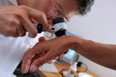
Skin examination
Medical expert of the article

Last reviewed: 04.07.2025
The main complaint of patients, which makes them pay attention to the condition of the skin, is itching. More often it occurs simultaneously with skin changes that are detected during examination (for example, with psoriasis). However, itching can be a secondary manifestation of a disease of the internal organs, which is observed, for example, with diseases of the liver and bile ducts, lymphogranulomatosis. Itching can also be the first sign of intolerance to a drug, other allergic conditions (urticaria in response to certain foods, etc.). Skin itching can be so unbearable that sleep-deprived patients resort to the most extreme measures, suicidal attempts are possible. With prolonged severe itching, traces of scratching are usually found on the skin.
Visual inspection of the skin
Skin color changes may be local or widespread. Pallor and redness of the skin may alternate due to fluctuations in blood supply in people with a labile autonomic nervous system. Constant and most often increasing pallor is observed with a decrease in the hemoglobin content in the blood (anemia ), for example, with acute blood loss or with various blood diseases. Whitening with a feeling of numbness, for example, of the fingers (the "dead fingers" symptom) is observed with vascular spasms - Raynaud's disease. The skin and mucous membranes may acquire a bluish tint (cyanosis) in heart failure with blood stagnation in the systemic circulation and an increase in the content of reduced hemoglobin in the blood due to this. The skin acquires a peculiar pale coffee shade (the color of “coffee with milk”) in case of untreated subacute infective endocarditis; in case of uremia, the skin color is pale greenish (anemia with retention of urochromes in the skin).
Jaundice of the skin is observed in diseases of the liver and bile ducts as a result of the accumulation of bilirubin in the blood (hyperbilirubinemia), which can also form in large quantities during the breakdown of red blood cells (hemolysis). Jaundice first appears on the sclera, then spreads to the mucous membrane of the oral cavity, the skin of the palms, and other areas. With prolonged severe hyperbilirubinemia, jaundice can acquire a greenish or dark ("earthy") hue.
With adrenal insufficiency, the skin appears tanned, which is also observed in hemochromatosis (iron retention in tissues). Eating large amounts of certain foods (for example, carrots and tomatoes containing carotenes) or taking certain medications can also cause changes in skin color.
Loss of pigment in certain areas of the skin occurs in the form of vitiligo - depigmented white spots, often located in symmetrical areas.
The skin of the face takes on a characteristic appearance in many patients who abuse alcohol: the skin of the nose and cheeks has a purple-blue tint, and there is a marked dilation of the scleral vessels.
Various skin rashes are of great diagnostic importance. Thus, in a number of infectious diseases they often "reveal" the diagnosis, in other cases they help to differentiate the disease. Hemorrhages and small hemorrhagic (petechial) rashes occur in various pathologies and not only in connection with blood clotting disorders. Large-spotted reddening of the skin (erythema) have different origins. The so-called nodular erythema on the anterior surface of the shins with painful compaction of the erythematous area of the skin is observed most often in sarcoidosis, as well as in tumors, drug intolerance, tuberculosis. For example, hemorrhagic rash as a manifestation of hemorrhagic vasculitis is of great importance for diagnosis - primary ( Schonlein-Henoch disease ) and secondary (in chronic liver diseases, some tumors).
When examining the skin, you can detect trophic disorders, bedsores in areas that are subject to prolonged pressure, as well as moles, tumor formations ( basaliomas, rarer tumors, tumor metastases). You should pay attention to tattoos, which can cause hepatitis B and C viruses to enter the body, which helps to understand the etiology of the detected changes in the liver and other organs. Postoperative scars after opening abscesses and fistulas should also be recorded. Traces of small burns on the skin are often found in people suffering from alcoholism.
Various skin changes are observed in systemic diseases of connective tissue. In systemic lupus erythematosus, the appearance of erythematous rashes on the cheeks in the form of butterfly wings and the bridge of the nose is characteristic. In systemic scleroderma, a mask-like appearance of the face, the disappearance of facial expressions, and the appearance of folds around the mouth in the form of a purse string are noted.
Livedo (Latin: bruise) is a special skin condition characterized by its bluish color due to the mesh or tree-like pattern of vessels visible through the skin. The following types (stages) of livedo are distinguished:
- marbling of the skin;
- reticular livedo - livedo reticularis;
- tree-like livedo - livedo racimosa.
Livedo is most often observed in systemic lupus erythematosus, Sneddon's syndrome, nodular periarteritis, and can also be observed in other diseases: dermatomyositis, systemic scleroderma, infections (tuberculosis, malaria, dysentery ); a connection with hyperproduction of antibodies to phospholipids (cardiolipin, phosphatidylserine) has been noted, and the pathogenetic significance of the latter in the development of livedo is discussed.
Xanthomas appear as whitish spots that rise above the surface of the skin and are associated with cholesterol deposits.
Peculiar dilation of skin vessels ( telangiectasias ) in the form of “spider veins” are observed in chronic liver diseases ( liver cirrhosis ).
Skin hypersensitivity reactions (allergies) to various substances, primarily to medications and food products, can manifest themselves as various rashes and itching, such as so-called urticaria.
Rapid hair loss is observed, for example, in systemic lupus erythematosus. Hirsutism, i.e. excessive hair growth on the face, trunk, legs, is observed in young women as a result of excess circulating androgens (male sex hormones). In hypogonadism, i.e. decreased function of the sex glands, both men and women experience insufficient hair growth in the armpits and pubis.
Nails can change with various diseases. The most well-known changes are those of the terminal phalanges of the fingers, which take on the appearance of so-called drumsticks, with the nails appearing convex, like watch glasses (Hippocratic fingers). Similar changes are observed in chronic suppurative diseases ( bronchiectasis, osteomyelitis ), as well as in infective endocarditis, chronic liver disease, and congenital heart defects. With prolonged iron deficiency, nails can become flat and then spoon-shaped (koilonychia).
Palpation of the skin
Palpation of the skin is usually performed together with its inspection. In this case, increased dryness or, conversely, moisture of the skin can be detected. Excessive sweating is observed with an increase in body temperature, hyperthyroidism, hypoglycemia, hot flashes in women during menopause, some nervous conditions, pain, fear. Increased sweating is observed with tuberculosis, infective endocarditis. Sometimes with increased sweating, a special rash ( prickly heat ) appears in the form of bubbles the size of a poppy seed, covering the skin like dew. Sweat contains most of the electrolytes of the blood, but in a concentration significantly lower than plasma. Sometimes, studying the composition of sweat can provide valuable diagnostic information. Thus, with cystic fibrosis, a characteristic increase in the concentration of sodium chloride in sweat is noted, with diabetes mellitus, the glucose content in sweat increases, which predisposes to skin infection ( furunculosis ).
Palpation may also reveal a local increase or decrease in skin temperature. An increase in temperature may be associated with increased blood flow during inflammation, such as erysipelas, or deep vein thrombophlebitis of the shins. A decrease in the temperature of the extremities is observed when arterial blood flow is disrupted.
When palpating the skin, its elasticity or resilience is determined, which depends on the state of its colloids and blood supply. With aging and some chronic diseases with fluid loss, the elasticity of the skin decreases, it is easy to take into a fold. The elasticity of the skin is especially sharply reduced in Ehlers-Danlos disease.
 [ 4 ], [ 5 ], [ 6 ], [ 7 ], [ 8 ]
Skin appearance assessment
Visual analysis of the skin is important for primary diagnostics and for testing cosmetic products. Today, there are several types of systems that allow for a quick and comfortable objective assessment of the skin's appearance. The assessment is based on two main parameters:
- surface topography and
- skin color.
The method of casts (method of replicas)
A skin cast is obtained by applying a thin layer of a special compound (nitrocellulose or silicone) to it. The compound hardens to form a film, the inner surface of which replicates the skin surface. After the cast is taken, the relief surface is scanned, the data is digitized and analyzed. This is how the wrinkle density (the number of wrinkles per unit of surface), their area and depth are determined. The surface of the cast is enlarged under a microscope and photographed; the resulting gray image is digitized and the parameters characterizing the skin relief are calculated.
[ 12 ], [ 13 ], [ 14 ], [ 15 ]
Direct optical scanning of the skin
Just a few years ago, the cast method was probably the most common way to study skin topography. However, it has a number of inconveniences, primarily related to the production of the cast itself. Therefore, the search for alternative methods has not ceased. Today, a new generation of technologies is coming to replace them - contactless, fast, safe and accurate, performing direct optical scanning of the skin surface.
Modern devices are equipped with powerful computers with specially developed software that allows, among other things, editing three-dimensional color images.
As an example, we will cite PRIMOS - a system for optical three-dimensional skin analysis, developed by the German company GFMessetechnik GmbH. The PRIMOS scanner is a complex optical device consisting of many micromirrors that "read" information from the scanned surface at different angles. The measuring accuracy of the scanner is impressive: it distinguishes points located at a distance from several millimeters to several micrometers from each other! PRIMOS takes a topographic image of the skin surface and, based on the resulting image, evaluates the relief, for example, determines the degree of roughness, "digitizes" wrinkles, scars, etc.
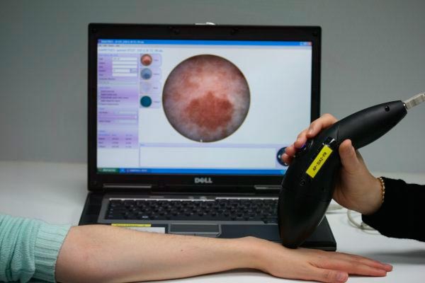
Another example of a direct scanning system is the SIAScope, an advanced method of dermatoscopy (Astron Clinica Ltd., UK). SIAScope obtains information about the skin's condition based on spectral analysis of light reflected from the skin's surface. To do this, SIAScope illuminates the skin with visible or near-red light that is safe for the body and then records the reflected light, sequentially obtaining 8 images at wavelengths from 450 to 950 nm (from blue to near-red). The resulting combined image is a circle 11 mm in diameter with a resolution of over 900 dots/mm 2. The image is then analyzed in accordance with the optical model of the skin, according to which skin color depends on the main chromophores - the pigments melanin and hemoglobin; the intercellular substance of the dermal layer, consisting mainly of collagen fibers, also contributes to the skin's tone. The initial dermoscopic image is decomposed by spectral characteristics into several so-called SIAgraphs, the analysis of which allows us to draw conclusions about the level of skin pigmentation, blood supply and the state of the dermal matrix. The method is highly sensitive and has been tested in a number of major clinics, where it has proven itself well, in particular, for express diagnostics of melanoma.