
All iLive content is medically reviewed or fact checked to ensure as much factual accuracy as possible.
We have strict sourcing guidelines and only link to reputable media sites, academic research institutions and, whenever possible, medically peer reviewed studies. Note that the numbers in parentheses ([1], [2], etc.) are clickable links to these studies.
If you feel that any of our content is inaccurate, out-of-date, or otherwise questionable, please select it and press Ctrl + Enter.
Dermatoscopy
Medical expert of the article

Last reviewed: 06.07.2025
Dermatoscopy is a modern method used to diagnose various neoplasms on the skin without the need for surgical intervention. Thanks to it, a specialist is able to record the changes that occur in the neoplasm at the early stages of degeneration. Thus, a doctor can see the development of a malignant tumor even before symptoms appear.
Advantages and disadvantages
The main advantages of this method include:
- Possibility of examination of moles and other neoplasms of any size, even the smallest.
- There is no need to damage the skin or the surface of the nevus.
- It is possible to diagnose melanoma at an early stage.
- Dermoscopy is a very quick procedure, it rarely takes more than half an hour.
- The specialist receives the result immediately.
Due to the fact that this method is completely safe and fast, it is quite difficult to find any disadvantages in it. The only thing is that when detecting melanoma, the doctor can be sure of the diagnosis only by 80%, so after this it is still necessary to conduct a histological examination.
Indications for the procedure
Dermatoscopy is used for:
- The appearance of pigmented nevi or moles.
- Conducting diagnostics of cancerous formations on the skin.
- Conducting diagnostics of keratoma or solar keratosis.
- The appearance of a hemangioma or angioma.
- Conducting diagnostics of papillomas and warts.
In what situations might a specialist recommend performing a dermatoscopy?
- If a new mole appears on the patient’s body, which is characterized by rapid growth, or an old mole begins to change (itch, peel).
- If the patient accidentally injured the nevus.
- The patient has decided to remove a raised mole on the face or another part of the body and wants to make sure that the growth is benign.
- If a person has a lot of moles and new nevi constantly appear.
- The patient had a family history of skin cancer/melanoma.
- A congenital nevus is located in a place where it is constantly rubbed by clothing and becomes inflamed from time to time.
Preparation
Since dermatoscopy is a visual diagnostic method, the patient does not need to be specially prepared for it. Neither anesthesia nor any serious preparation is used before this method. The only request: do not apply any creams or other cosmetics to the mole on the day of the examination.
 [ 9 ]
[ 9 ]
Who to contact?
The device for carrying out the procedure
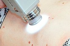
Dermatoscopy is performed using a special device called a dermatoscope, which allows you to magnify various skin lesions dozens of times and fully examine its entire structure. As a rule, it is most often used during melanoma diagnostics. Modern dermatoscopes not only magnify a mole, but also take digital photographs of the lesions, which allows you to display them on a screen and examine them more thoroughly.
In addition, the digital dermatoscope shines through the upper layers of the epidermis and helps to see what is happening with the neoplasm inside. The device contains a set of achromatic lenses, which are distinguished by a high degree of resolution.
What are digital dermatoscopes used for?
- Diagnosis of any changes in the skin, especially those that lead to the development of moles.
- They help to identify malignant neoplasms at an early stage.
- With their help, you can evaluate various moles.
- They allow an assessment of the skin structure.
The dermatoscope has such functionality that a specialist can easily make a correct diagnosis, evaluate a mole, the symmetry or asymmetry of the neoplasm, check the size and shade, the edges of the mole and the possible presence of dots inside it.
Today, manufacturers produce a huge number of different dermatoscopes. The most popular among them are the following models:
- HeineMini 3000 is a pocket-sized dermatoscope that runs on battery power (it can work for nine hours without recharging).
- HeineDelta 20 plus – has LED lighting, thanks to which the specialist can more effectively study the surface of the neoplasm.
- KaWePiccolightD – suitable for early diagnosis of melanomas.
- AramoSG – a dermatoscope that can be connected to a computer.
Technique dermatoscopies
During dermatoscopy, a specialist uses a dermatoscope to examine the entire surface of the skin neoplasm and adjacent layers.
Before the procedure, the patient must lie down or sit down, and also expose the area where the mole is located. In some cases, it is necessary to apply a little gel or special oil to the skin. They will help reduce the reflection on the skin and increase the effectiveness of the method.
Digital and computer dermatoscopy
Today, digital dermatoscopy is the most popular and effective method for diagnosing melanoma and other malignant skin tumors. It especially helps to identify melanoma at an early stage of the disease, when other methods are powerless. Thanks to digital dermatoscopy, a specialist can immediately determine whether the formation is benign or not.
The process of examining the skin with a digital dermatoscope is somewhat similar to an ultrasound examination. The dermatoscope is pressed against the surface of the nevus, illuminating it enough to make images of the internal structure of the mole. This image can then be displayed on a computer screen, which allows for an even better examination of the neoplasm. In just a couple of minutes, a specialist can examine all the changes that have occurred in the mole and make an accurate diagnosis.
Epiluminescent dermatoscopy
ECD or epiluminescent computer dermatoscopy is the most modern method of diagnosing neoplasms on the skin. Its main difference is the use of polarized lighting, which better illuminates the mole from the inside and allows the specialist to examine all its features more closely. Thanks to ECD, it is possible to diagnose skin cancer with an accuracy of up to 95%.
An oncologist, having received the data of epiluminescent dermatoscopy, can determine whether it is necessary to remove the nevus, how important surgical intervention is. In addition, all images are stored in the computer and if the patient visits the doctor again, the doctor can compare the old images with the new ones.
Contraindications to the procedure
The main advantage of dermatoscopy as the main method of diagnosing malignant neoplasms on the skin is the fact that it has no contraindications. It can be carried out without fear even during pregnancy or breastfeeding. The fact is that it is during this period that women can develop a very large number of new neoplasms and it is important to check them as early as possible to avoid skin cancer.
Normal performance
[ 20 ], [ 21 ], [ 22 ], [ 23 ]
Dermoscopy for melanoma
In melanoma, dermatoscopy is an inexpensive and effective diagnostic method. It is non-invasive and in the process the doctor uses a special instrument called a "dermatoscope". A liquid (alcohol or imperial oil) is applied to the neoplasm, due to which the structures located in the upper layer of the dermis can be easily visualized.
The doctor evaluates the structural components and color shades of the nevus, which allows for a quick differentiation of the non-melanocytic or melanocytic nature of the mole. This method can also detect some structural phenomena of the neoplasm. For example, if a patient has areas without structure, this means that the tumor (melanoma) is regressing. If small black dots appear inside the structure, this indicates that the neoplasm is malignant.
Dermatoscopy can help avoid complex surgical operations on pigmented lesions. This method allows increasing the accuracy of the diagnostic assessment several times. If you combine dermatoscopy and clinical diagnostics, you can increase the likelihood of timely detection of melanoma. In clinical diagnostics, specialists use the so-called "ABCD rule" (it stands for: asymmetry, boundaries, shade and size). If the doctor sees that the neoplasm meets this rule, then most likely it is melanoma.
Recently, more and more doctors are paying attention to digital dermatoscopy, which helps to see a full picture of a nevus. This method is especially indispensable in diagnosing melanoma at early stages of development.
Dermoscopy of basalioma
Basal cell carcinoma or basalioma is a fairly common type of skin cancer (76% of cases). The main difference of this tumor is its benign course. It is very important to detect basalioma at an early stage, since it is at this time that it does not yet metastasize and is easily treated.
Dermatoscopy is one of the most accurate methods for diagnosing basal cell tumors. It is distinguished by the fact that it is not traumatic and helps to quickly conduct the necessary examination of the skin. Thanks to dermatoscopy, the doctor can accurately determine basalioma, which, during clinical examination, is often confused with some other skin diseases: trichoepithelioma, psoriasis, dermatofibroma, dermatitis, syphilis, melanoma.
The main signs of basalioma that a specialist detects during a dermatoscopy are:
- Clearly visible homogeneous zones that are distinguished by a bright red or white color.
- Presence of small ulcers.
- The structure is dominated by branching capillaries.
[ 27 ], [ 28 ], [ 29 ], [ 30 ], [ 31 ], [ 32 ], [ 33 ], [ 34 ], [ 35 ]
Dermoscopy of nevus
When a new mole appears or an old nevus changes quickly, it is very important to immediately contact a specialist. He will perform a dermatoscopy, which will help to detect skin cancer in time and carry out the necessary treatment. The main feature of this method in diagnosing a nevus is the fact that it helps to see even the most insignificant changes. Thanks to dermatoscopy, the doctor conducts the most thorough and accurate analysis of the nevus. This is explained by the fact that the dermatoscope allows you to enlarge the neoplasm up to ten times.
What does a doctor pay attention to when examining a patient with a dermatoscope? First of all, the size, symmetry, shade, border and structure of the nevus are examined. If it is a benign formation, then its appearance will always be symmetrical. If even a slight asymmetry appears, we can talk about the development of skin cancer.
Dermoscopy of a nevus is 80% accurate and does not require any special preparation from the patient for the procedure. If you have moles that bother you, it is important to have a dermatoscopy at least once a year.
[ 36 ], [ 37 ], [ 38 ], [ 39 ], [ 40 ], [ 41 ], [ 42 ], [ 43 ], [ 44 ]