All iLive content is medically reviewed or fact checked to ensure as much factual accuracy as possible.
We have strict sourcing guidelines and only link to reputable media sites, academic research institutions and, whenever possible, medically peer reviewed studies. Note that the numbers in parentheses ([1], [2], etc.) are clickable links to these studies.
If you feel that any of our content is inaccurate, out-of-date, or otherwise questionable, please select it and press Ctrl + Enter.
Retina can help track the development of multiple sclerosis
Medical expert of the article
Last reviewed: 01.07.2025
Scientists at Johns Hopkins University in Baltimore have found that a routine eye exam can quickly and easily provide a health assessment for people with multiple sclerosis.

At the moment, there is no medicine that can stop the disease; the most that can be done is to slow down the progression of the disease.
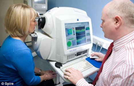
A new method for diagnosing multiple sclerosis is called optical coherence tomography, which is used in ophthalmology. It can be performed at a doctor's office and takes only a few minutes.
This technique allows tracking disease processes in patients with multiple sclerosis by the thickness of the retina, and the degree of its thinning will accurately tell doctors the speed at which the disease is progressing.
The secondary sign of an autoimmune disease is damage to neurons in the brain and spinal cord, and the primary sign is the destruction of myelin. Accordingly, for the timely detection of multiple sclerosis, it is necessary to examine tissues that are deprived of the myelin sheath, for example, the inner shell of the eye - the retina.
The experiment by scientists, led by Doctor of Medical Sciences and lead author of the study Peter Calabresi, involved 164 people – people suffering from multiple sclerosis, as well as 59 completely healthy people who were included in the control group. For 21 months, every six months, they underwent eye scanning using optical coherence tomography. At the beginning of the experiment and then every year, they also underwent magnetic resonance imaging of the brain.
The researchers found that patients with relapsing-remitting MS (a form in which symptoms disappear for a time) had 42% faster retinal thinning than others. Those with active inflammation, known as gadolinium lesions, had 54% faster retinal thinning. Those with T2 lesions had 36% faster retinal thinning.
In addition, the experts noted that in those patients whose disability worsened over the entire study period, the retina became 37% thinner compared to those who showed no signs of deterioration.
Retinal thickness decreased 43% faster in patients who had been sick for less than five years compared to those who had been sick for a longer period of time.
The study findings suggest that in people with a shorter duration of the disease and a more active form, retinal thinning may progress at a more rapid rate.
Multiple sclerosis is a progressive disease of the nervous system, which, despite its seemingly self-explanatory name, has nothing in common with absent-mindedness or senile sclerosis. The name of the disease is due to the peculiarity of the location of sclerosis foci throughout the nervous system, which replace nervous tissue with connective tissue.

