
All iLive content is medically reviewed or fact checked to ensure as much factual accuracy as possible.
We have strict sourcing guidelines and only link to reputable media sites, academic research institutions and, whenever possible, medically peer reviewed studies. Note that the numbers in parentheses ([1], [2], etc.) are clickable links to these studies.
If you feel that any of our content is inaccurate, out-of-date, or otherwise questionable, please select it and press Ctrl + Enter.
Chloasma spots on the face, body, arms and legs
Medical expert of the article

Last reviewed: 12.07.2025
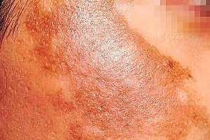
Chloasma is hyperpigmentation of a limited area of facial skin. It manifests itself as the appearance of brownish pigment spots. This is an acquired disease caused by excessive accumulation of melanin in the upper layers of the skin.
 [ 1 ]
[ 1 ]
Causes chloasmas
To date, there is no reliable information regarding what exactly is the root cause of chloasma. This pathological condition is associated with a violation of the melanin pigment metabolism. Most likely, this occurs due to endocrine and hormonal disorders in the body.
[ 5 ]
Risk factors
There are risk factors that can contribute to the development of chloasma:
- female gender;
- pregnancy;
- presence of hormonal disorders;
- diseases of the female reproductive system;
- prolonged exposure to UV rays, in particular, frequent visits to solariums, prolonged exposure to the sun;
- genetic predisposition, that is, the presence of relatives with chloasma in the family history;
- chronic liver diseases;
- taking oral contraceptives;
- hormone-producing tumors;
- gastrointestinal pathology;
- hypo- or avitaminosis;
- metabolic disorders;
- helminthic invasion;
- malaria;
- tuberculosis;
- damage to the epidermis as a result of careless squeezing of pimples
- the use of cosmetics that are not suitable for a particular skin type, are of poor quality and have a negative impact on the skin;
- hormone-containing therapy;
- CNS diseases;
- pathology of the endocrine system.
Symptoms chloasmas
Chloasma is manifested by the appearance of a hyperpigmented area of skin that has limited uneven edges. It has no elevation above the skin. Each person's chloasma can have its own shade with a predominant brown color. The size of the area of increased pigmentation can vary: from a few millimeters to a large affected area.
Pigmentations are solitary, but if they are located at a minimum distance from each other, the impression of multiple lesions may be created. Pain and itching are not typical for them. Patients may experience aesthetic discomfort.
The diagnostic sites where the symptoms of chloasma most often appear are the forehead, the area around the eyes, the nose, the upper lip, and the cheeks. Also, in rare cases, hyperpigmentation can be seen on the chest, back, midline of the abdomen, and inner thighs, on the legs.
Forms
There are several types of chloasma. One of them is perioral chloasma, which is diagnosed in females. It manifests itself as symmetrically located brown spots around the mouth. This type of chloasma has a long course, over time the saturation of the spots may change, and areas of hyperpigmentation may appear on the nasolabial folds.
The pigment line is also considered a type of chloasma. This is a dyschromic form that can be recognized by the appearance of a pigmented stripe about 10 mm wide, which is localized on the forehead, passes through the cheek to the outer side of the neck. However, this is a precursor to serious diseases associated with dysfunction of the nervous system, such as: brain tumor, parkinsonism, meningovascular syphilis.
Chloasma may be a manifestation of diseases of internal organs. For example, with cirrhosis, hepatitis, functional and organic liver damage, dysfunction of the bile ducts, so-called hepatic chloasma may occur, which is a reason to see a doctor and undergo a medical examination.
The skin is a hormone-dependent organ, so chloasma is often diagnosed as being caused by taking oral contraceptives, which change a woman’s hormonal background and cause a disruption in melanin metabolism.
Chloama of pregnant women also occurs against the background of hypersensitivity of the woman's skin to the effects of UV rays due to increased levels of estrogens in the body. It is characterized by specific areas of damage - nipples of the mammary glands and external genitalia. It is believed that hyperpigmentation during pregnancy does not require treatment. If the initial cause of the appearance of pigment spots is pregnancy, then after childbirth they should disappear.
Chloasma in children is diagnosed extremely rarely.
Diagnostics chloasmas
Chloasma diagnostics is based on several types of examination. First, the doctor must evaluate the appearance of the affected skin area, collect a history of life and disease, clarify whether this pigment spot is congenital or acquired, and check for the presence of an inflammatory process.
Specific diagnostic methods are prescribed. These are dermatoscopy (examination of the hyperpigmented area of skin using a dermatoscope, which allows for a tenfold increase in the field of vision) and siascopy (examination using a siascanner, which allows for the microscopic structure of pigment-containing cells to be seen), anda skin biopsy may be prescribed.
After this, laboratory tests begin. The patient takes a general blood test, general urine test, biochemical blood test, and coprogram. To exclude the liver type of disease or chloasma caused by diseases of the internal organs, biochemical liver tests, dysbacteriosis analysis, gastroscopy, ultrasound of the abdominal organs and liver are prescribed. For women, a gynecologist's examination is required to exclude pathology associated with dysfunction of the reproductive system.
Differential diagnosis
When diagnosing chloasma, differential diagnostics are carried out with various diseases caused by a disorder of melanin metabolism.
For example, a pigment spot is also a skin area with increased pigmentation. However, depending on the type of pigment spot, it can have a smooth outline, appear on any part of the body, unlike chloasma, which appears as a brown spot with uneven borders and has favorite places on the body where it most often appears.
Chloasma and lentigo also have some similarities. Lentigo is a skin disease that is most often diagnosed in people over 40. The spots are round or oval in shape, can rise above the skin level, and are a risk factor for the development of tumor-like diseases.
Who to contact?
Treatment chloasmas
In order for the treatment to give a positive result, it is necessary to find out the initial cause that provoked the development of this disease. For example, if chloasma is a consequence of hormonal imbalance, drugs are prescribed to correct this condition, or oral contraceptives are changed if this was the cause. Treatment of liver chloasma includes hepatoprotectors and drugs necessary to restore liver dysfunction.
Other treatment methods are also distinguished.
Laser or chemical peeling removes the top layer of skin. This method is effective only for shallow spots.
Fractional or neodymium lasers are used to treat chloasma. The first removes cells with increased melanin content, affecting the surrounding tissues, the second is more gentle in this regard.
Photocorrection is carried out by exposing the skin to high-density light pulses, as a result of which the melanin-containing pigment is destroyed.
Mesotherapy is one of the effective methods of treating chloasma, in which a solution containing useful vitamin complexes, such as ascorbic and glycolic acids, is injected into the skin. They help suppress the activity of cells containing melanin and destroy it.
Whitening ointments and creams, which include inhibitors of melanin precursor, inhibitors of coloring pigment formation, such as: hydroquinone, azelaic acid, arbutin. Examples can be various ointments: 5% hydroquinone ointment, Achromin, Melan.
Vitamin therapy includes taking folic acid, ascorbic acid, B vitamins, and riboflavin.
There are a number of folk recipes for treating chloasma at home:
- applying a mixture of hydrogen peroxide and lemon juice to the hyperpigmented area with a cotton swab;
- gauze soaked in milk is placed on chloasma for 20 minutes;
- wiping the skin with chamomile infusion;
- Using a cotton pad, apply a decoction of parsley and lemon juice to the affected skin for 20 minutes.
Prevention
Prevention of chloasma includes:
- avoiding prolonged exposure of the skin to sunlight, especially in the summer;
- use of sunscreens;
- use natural sun protection measures: hat, bandana, panama, cap, sun umbrella, glasses;
- avoid wearing clothing that may cause chafing;
- avoid contact with chemicals that have a negative effect on the skin (gasoline, machine oil);
- carefully select oral contraceptives after consulting a gynecologist;
- do not use low-quality cosmetics that do not suit your skin type;
- undergo an annual medical examination to prevent the development of internal organ diseases;
- have a healthy, nutritious diet, replenishing the body with a sufficient amount of vitamins, electrolytes, and beneficial metabolites.
Forecast
The prognosis for the person's life and work activity is favorable.
[ 27 ]