
All iLive content is medically reviewed or fact checked to ensure as much factual accuracy as possible.
We have strict sourcing guidelines and only link to reputable media sites, academic research institutions and, whenever possible, medically peer reviewed studies. Note that the numbers in parentheses ([1], [2], etc.) are clickable links to these studies.
If you feel that any of our content is inaccurate, out-of-date, or otherwise questionable, please select it and press Ctrl + Enter.
Hyperpigmentation of the skin
Medical expert of the article

Last reviewed: 07.07.2025
Pathogenesis
Histological examination of the skin reveals hyperkeratosis with horny plugs in the mouths of hair follicles, sometimes atrophic changes in the epidermis and vacuolar degeneration of the cells of the basal layer. In the dermis, as a rule, there is a varying degree of inflammatory reaction, in the cells of the basal layer of the epidermis, as well as in melanocytes, the content of melanin is increased, especially large amounts of it are found in the cytoplasm of macrophages of the upper third of the dermis, or melanophages. The superficial capillaries are dilated, which is clinically manifested by telangiectasias. Small infiltrates are visible around them, consisting mainly of lymphocytes with an admixture of tissue basophils.
 [ 8 ], [ 9 ], [ 10 ], [ 11 ], [ 12 ], [ 13 ], [ 14 ], [ 15 ]
Symptoms skin hyperpigmentation
Limited hyperpigmentation includes freckles, chloasma, café-au-lait pigment spots, simple and senile lentigo, Becker's nevus, iatrogenic melanosis and post-inflammatory hyperpigmentation.
Freckles are small (2-4 mm), pigmented spots of tan color with unclear outlines. They appear at any age on exposed areas of skin, especially in fair-haired and white-skinned people, darken under the influence of sunlight, and disappear in winter.
Pathomorphology. Hyperpigmentation of epidermal cells, especially the basal layer, is determined. There is no melanocyte proliferation.
Histogenesis. Under the influence of ultraviolet radiation, there is an increase in the synthesis of melanin in the epidermis and its accumulation in melanocytes and keratinocytes.
Chloasma is a larger pigmented spot that occurs due to liver dysfunction, endocrinopathies, pregnancy and diseases of the appendages in women.
Pathomorphology. Increased melanin content is noted in epidermal cells.
Lentigo simplex is a spotted element with a diameter of 1 to 3 mm, with clear contours, dark brown or black in color. It appears at any age, including childhood, on open areas of the body.
Pathomorphology. The number of melanocytes in the basal layer of the epidermis is increased, but unlike the border nevus, they do not form "nests". At the same time, melanocytes usually increase in size. At the same time, there is an increase in the number and lengthening of epidermal outgrowths (lentiginous hyperplasia of the epidermis). The content of melanin in the basal layer is increased. In the dermis - small lymphocytic infiltrates and single melanophages.
Histogenesis. Skin hyperpigmentation is based on local proliferation of melanocytes.
Widespread lentiginous hyperpigmentation is observed in xeroderma pigmentosum, periorificial lentiginosis.
Xeroderma pigmentosum is a heterogeneous, predominantly autosomal recessive disease characterized by increased photosensitivity, development of pigmentation and skin atrophy, photophobia, neurological symptoms, progressive course with a very high risk of developing skin tumors. Increased sensitivity of cells to ultraviolet rays is due to impaired DNA reparation, and insufficiency of endonuclease excision of pyrimidine dimers is possible. Some patients experience neuropsychiatric symptoms and hypogonadism (de Sanctis-Cacchione syndrome).
Pathomorphology. The histological picture at the initial stage of the disease is nonspecific. Hyperkeratosis, thinning of the Malpighian layer of the epidermis with atrophy of some epithelial cells and an increase in the volume of others, accompanied by uneven accumulation of melanin in the cells of the basal layer and an increase in the number of melanocytes are noted. A small lymphocytic infiltrate is visible in the dermis. In the stage of hyperpigmentation and atrophic changes, hyperkeratosis and pigmentation are more pronounced. The epidermis is atrophic in some areas and thickened in others. There is a violation of the arrangement of the nuclei of the epithelial cells, an increase in their volume, atypical forms appear, as a result of which the picture resembles solar keratosis. In the dermis - dystrophic changes similar to those in solar dermatitis, characterized by basophilia of collagen fibers and elastosis. In the later stages of the disease, atypical growths of the epidermis join the changes described above, and in some areas squamous cell carcinoma and sometimes basal cell carcinoma develop.
Lentiginosis periorificialis (syn.: Peutz-Jeghers-Touraine syndrome) is a neuromesenchymal dysplasia caused by a gene mutation transmitted in an autosomal dominant manner. The disease develops in the first years of life, but can exist from birth, and rarely occurs in adults. Clinically, multiple, small pigmented spots from light brown to black, oval or rounded in shape, densely located around the mouth, on the lips, especially the lower one, perinasal, periorbital and on the mucous membrane of the oral cavity are detected. Less often - on the extremities (palms, soles, dorsum of the fingers). A.V. Braitsev and G.M. Bolshakova (1960) described generalized lentiginous rashes. Periorificial lentigo is combined with intestinal polyposis, mainly of the small intestine, prone to transformation into adenocarcinoma.
Pathomorphology. An increase in the amount of pigment in the cells of the basal layer is noted, accompanied by an increase in the number of melanocytes. In the upper parts of the dermis, a large number of melanophages are found, the pigment melanin is sometimes located extracellularly.
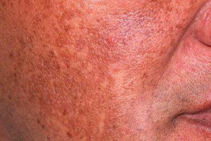
Lentigo senile (syn.: solar lentigo) appears in middle-aged and elderly people after repeated exposure to ultraviolet radiation, especially after sunburn. The preferred localization is open areas of the body, the skin in the shoulder girdle and upper back. The size of the lentiginous elements is from 4 to 10 mm, the color is from light brown to dark brown and even black, the outlines are blurred, uneven,
Pathomorphology. Lentiginous hyperplasia of the epidermis, hyperpigmentation of keratinocytes of the basal layer, minor proliferation of melanocytes. In the dermis - dystrophic changes in collagen fibers, manifested by their basophilia (solar elastosis).
Café-au-lait spots are large pigmented yellowish-brown spots that are congenital or appear soon after birth. Their surface is smooth, and their outlines are often oval. With age, the number and size of the spots increase. Multiple spots are pathognomonic for neurofibromatosis, and are observed in other genodermatoses, such as tuberous sclerosis and Albright's disease, but single elements can also be found in healthy individuals.
Pathomorphology. Hyperpigmentation of the basal layer of the epidermis, giant granules (macromelanosomes) are detected in DOPA-positive melanocytes.
Becker's nevus (syn.: Becker's neviform melanosis) is a local skin lesion, usually in the shoulder girdle area, manifested by a hyperpigmented area of a rich brown color, usually in combination with pronounced hypertrichosis within the nevus. It is a developmental defect, observed mainly in males, the full clinical picture develops in adolescence, pigmentation increases under the influence of ultraviolet rays.
Pathomorphology. Hyperpigmentation of the basal layer, acanthosis and hypertrichosis. Often observed in combination with underlying smooth muscle hamartoma, changes in collagen fibers in the nevus area have been described, which gives grounds to consider it an organoid nevus.
Secondary hyperpigmentation appears in places of primary morphological elements of the rash - papules, tubercles, vesicles, pustules, as well as secondary elements - erosions and ulcerative lesions, after an acute or chronic inflammatory process. This type of pigmentation is based on an increase in the amount of pigment in the cells of the basal layer of the epidermis and melanocytes, which remains after the inflammation disappears.
Pathomorphology. An increase in pigment content is noted in the basal layer, the thickness of which may vary depending on the nature of the former element.
Forms
Hyperpigmentation can be widespread and limited, congenital and acquired.
Widespread acquired hyperpigmentation of the skin is observed in cachexia due to debilitating diseases (cancer, tuberculosis, etc.), in vitamin deficiencies (pellagra, scurvy), and adrenal pathology (Addison's disease).
Frequently encountered skin diseases that occur with increased melanogenesis are melasma, which develops on the basis of intoxication, mostly of a professional nature (contact with flammable and lubricating substances). These include Riehl's melanosis, or reticular poikiloderma of Civatte, toxic melasma of Habermann-Hoffmann. In this case, the skin of the face, neck, chest and back of the hands is affected, clinically characterized by bluish-brown, generalized or limited, diffuse or reticular pigmentation.
What do need to examine?
How to examine?