
All iLive content is medically reviewed or fact checked to ensure as much factual accuracy as possible.
We have strict sourcing guidelines and only link to reputable media sites, academic research institutions and, whenever possible, medically peer reviewed studies. Note that the numbers in parentheses ([1], [2], etc.) are clickable links to these studies.
If you feel that any of our content is inaccurate, out-of-date, or otherwise questionable, please select it and press Ctrl + Enter.
Trachoma
Medical expert of the article

Last reviewed: 05.07.2025
Trachoma is a specific, contact-transmitted chronic infectious, usually bilateral, inflammation of the conjunctiva of the eye, expressed by its diffuse infiltration with the formation of follicles (grains), their degeneration, decay and subsequent scarring.
Epidemiology
Currently, trachoma affects about 400 million people worldwide, and there are 4 to 5 million people who are blind from trachoma. It is found primarily in Africa, the Middle East, Asia, Central and South America, especially in areas with overcrowding and poor sanitation.
Causes trachomas
The causative agent of trachoma is Chlamydia trachoma A, B, C, discovered in 1907 by Prowazek and Halberstadter. Chlamydia are obligate intracellular parasites. Trachoma is transmitted from eye to eye through contaminated hands or shared objects (towel). Flies also play an important role in the transmission of infection.
The incubation period of trachoma lasts from 5 to 12 days. The main essence of the conjunctival disease in trachoma is the formation of follicles and infiltration, a characteristic feature is the inevitable development of scars in the conjunctiva at the site of infiltration and follicles for typical trachoma. The disappearance of infiltration and the transformation of follicles into scar tissue ends trachoma. Trachoma affects only the conjunctiva of the eye and is not localized on other mucous membranes. In an experimental study of trachoma on animals, it was not possible to obtain typical trachoma on the conjunctiva of even anthropoid apes.
Symptoms trachomas
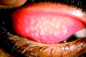
Trachoma is chronic. It usually begins unnoticed, with barely noticeable mucopurulent discharge from the conjunctival cavity, sometimes accompanied by itching, photophobia, lacrimation, pseudoptosis (due to swelling of the eyelids). The process is usually bilateral, more pronounced on the conjunctiva of the upper transitional fold of the upper eyelid.
The symptoms vary depending on the degree of infiltration, grains and papillae, and associated complications. However, it is possible to divide the course of trachoma into 4 stages.
Trachoma is characterized by the spread of the process to the cornea. In the superficial layer of the upper edge (limbus) of the cornea, small point infiltrates appear, to which thin loops of conjunctival vessels approach. In this case, patients experience lacrimation, photophobia, blepharospasm. Initial signs of corneal trachoma can appear already in its earliest stages, which is of great importance in diagnostics. Often, especially with early treatment, the damage to the cornea by trachoma can be limited to this. Then the infiltrates resolve, the eyes calm down, but the network of thin superficial vessels remains for life.
In more severe cases, a number of new infiltrates may appear, but below the place where the vessels have grown. Infiltrates may spread down the corneal rim, merge with each other, thus forming a diffuse superficial corneal opacity, which is penetrated by vessels. The corneal epithelium above the opacity becomes uneven and rough. Such superficial vascular inflammation of the cornea is called pannus (from the Greek pannus - "curtain").
Usually pannus, descending down the cornea, reaches its center and breaks off abruptly, but can spread further to the entire cornea. The degree of corneal infiltration and the development of vessels in it with pannus are very different. There are 2 forms of pannus: thin pannus, in which there is insignificant and barely expressed vascularized infiltration of the cornea; vascular pannus, in which the cornea, due to significant infiltration and an abundance of newly formed vessels, takes the form of fleshy growths and is therefore also called "sarcomatous pannus".
Trachomatous pannus occurs at any stage of trachoma, regardless of the severity and extent of the process in the conjunctiva. Trachomatous pannus may occur through the affected conjunctiva of the eyelids with the oral membrane or as a result of the spread of the process of the conjunctiva of the eyeball to the cornea. Trachomatous pannus, depending on its prevalence, nature and degree of changes in the cornea, reduces vision. Pannus has a high tendency to recur. Damage to the cornea is an almost constant companion of trachoma and serves as an important differential diagnostic sign, especially in the initial stage, when there are no signs of scarring. Therefore, if trachoma is suspected, the upper part of the limbus should be very carefully examined with a magnifying glass.
As already noted, in most cases trachoma begins unnoticed and develops gradually and slowly. Often patients, without experiencing any particular suffering, do not seek medical help for a long time, not knowing what the disease threatens them with in the future. At the same time, patients are a source of infection for others. Often such patients seek help only when they have purulent discharge from the eyes or when they begin to lose their vision.
Patients who seek help at the very beginning of the disease, when the initial forms of trachoma described above can be seen, complain of a feeling of a foreign body in the eye, heat, burning, the appearance of mucous discharge in the morning and glued eyelashes.
In contrast, some patients, despite the presence of signs of blooming trachoma and even an advanced scarring process, do not experience any unpleasant sensations. These patients are identified during preventive examinations of certain groups of the population, especially schoolchildren, since trachoma in children usually proceeds much more easily than in adults. The question of the possibility of an acute onset of trachoma, when the disease begins with acute inflammatory phenomena in the presence of photophobia, lacrimation, sharp pain and a large amount of purulent discharge, is controversial; then all these acute phenomena disappear, and follicles and infiltration, i.e. signs of the first stage of trachoma, come to the fore. Then the disease proceeds in its usual chronic form. A number of scientists categorically deny the possibility of acute trachoma, believing that in these cases some concomitant infection joins ordinary trachoma (Koch-Wilks bacilli, very common in trachoma, pneumococci, etc.).
Stages
The first stage of trachoma in the initial phase has a pronounced infiltration of the mucous membrane of the eyelids and the development of follicles only in the transitional folds: in the developed form, diffuse infiltration and follicles spread to the cartilage, especially to the upper eyelid. All phenomena gradually increase, but signs of scarring are completely absent. The first stage of trachoma can exist for months, years.
The second stage of trachoma is the further development of mature juicy follicles that look like stale raspberries; pannus and infiltrates in the cornea; the appearance of individual conjunctival scars due to follicle necrosis. However, at this stage, the phenomena of hypertrophy prevail over the phenomena of scarring; patients at this stage are most dangerous as a source of new infections, since overripe follicles are easily covered and their contents flow out. With a gradual decrease in inflammation (hyperemia, follicle infiltration) and an increase in scarring, the trachomatosis process passes into the third stage.
The third stage of trachoma is widespread conjunctival scarring with a combination of residual inflammatory infiltration and follicles. In the cicatricially altered conjunctiva, individual areas of redness and infiltration are still visible. The third stage of trachoma lasts for a long time and can often be accompanied by exacerbations of the inflammatory process and complications. At this stage, the consequences of trachoma already make themselves known.
The fourth stage of trachoma is the final scarring of the conjunctiva without inflammatory processes: hyperemia and visible infiltration. The conjunctiva has the appearance of a whitish, tendon-like surface, since it is completely or partially replaced by scar tissue in the form of a mesh and small strokes. The fourth (scarring) stage of trachoma determines clinical recovery (but the presence of deep infiltration is not always easy to exclude). This stage of trachoma is not contagious, unlike the first three, which can last for years.
Complications and consequences
The consequences of trachoma are varied. Replacement of infiltrates and follicles by connective tissue leads to cicatricial degeneration of the conjunctiva, as a result of which the transitional folds are shortened; the vaults are reduced or destroyed, which limits the movement of the eyeball. When pulling down the eyelid, especially the lower one, one can notice how the conjunctiva is stretched in the form of vertical folds (symblepharon).
The cicatricial change in the thickness of the cartilage and conjunctiva leads to contraction and, as a result, to a trough-shaped curvature of the cartilage, which subsequently causes the inversion of the eyelids. In this case, the ciliary edge of the eyelid facing the cornea constantly irritates and injures it.
Along with the inversion, and sometimes independently, trichiasis occurs - an incorrect position of the eyelashes. The eyelashes - all or part of them - are directed towards the eyeball when blinking, rubbing the cornea, causing irritation. The development of trichiasis is associated with the spread of trachoma to the edge of the eyelid, when the inflammatory infiltration is replaced by connective tissue and scars disrupt the correct position of the hair follicles. Scarring of the edge of the eyelids also leads to the closure of the excretory ducts of the meibolic glands, their cystic stretching and thickening of the cartilage.
With widespread conjunctival scarring, its glandular apparatus dies, the excretory ducts of the lacrimal glands close, the moistening of the conjunctiva and cornea decreases or stops, their sensitivity decreases, and metabolic processes are sharply disrupted. As a result, separate matte-white dry plaques appear on the conjunctiva; the same plaques form on the cornea, its epithelium becomes thicker, keratinizes, and acquires the character of the epidermis. The cornea becomes cloudy, opaque, and vision is sharply reduced. This condition is called deep parenchymatous xerosis.
The course of chronic trachomatosis can be complicated by acute inflammatory processes in the conjunctiva, cornea and lacrimal organs.
Acute infectious conjunctivitis is a common complication of trachoma and is caused by microorganisms such as Koch-Weeks bacillus, pneumococcus, and gonococcus.
Infections superimposed on the trachomatosis process aggravate its course and change the picture of trachoma, creating difficulties in its diagnosis. Complication of trachoma with acute conjunctivitis contributes to the spread of trachoma and is a great danger to the cornea.
A severe complication of trachoma is corneal ulcers. In some cases, this is a typical trachoma ulcer, in other cases, the ulcer develops at some distance from it on any part of the cornea. Ulcers can spread in width and depth and sometimes lead to a perforation of the cornea at the site of the ulcer, subsequently forming a dense opaque leukoma, causing a sharp decline in vision and often blindness. The development of ulcers is facilitated by friction of eyelashes on the cornea and eversion of the eyelids, which often occurs with trachoma.
Often, chronic inflammation of the lacrimal sac occurs with trachoma, as a result of which the lacrimal passage from the conjunctival sac into the nasal cavity is disrupted and panic conjunctivitis develops. This has an adverse effect on the course of trachoma.
The course of trachoma is long. It lasts for months, years, sometimes a lifetime. The general condition of the organism and its reactivity are of primary importance in the course of trachoma. Trachoma becomes more persistent and is difficult to treat in those who suffer from such general diseases as tuberculosis, scrofulosis, malaria, and helminthic invasion. General diseases, reducing the reactivity of the organism, aggravate the course of trachoma.
Trachoma is milder and less noticeable in children. It is in children that cases of spontaneous healing without particularly severe changes in the conjunctiva are more often observed.
Diagnostics trachomas
The diagnosis of trachoma is based on the characteristic clinical picture and laboratory test data, such as the predominance of polymorphonuclear leukocytes in conjunctival scrapings, the detection of intranasal inclusions (Prowazek-Halberstadter bodies) in the epithelial cells of conjunctival scrapings, and the detection of chlamydial particles in conjunctival scrapings by immunofluorescence using monoclonal antibodies.
 [ 21 ]
What do need to examine?
How to examine?
Who to contact?
Treatment trachomas
Chemotherapy consists of long-term local and general use of antibiotics and sulfonamides, which act on the causative agent of trachoma and eliminate the accompanying bacterial flora. For trachoma, two methods of treatment are used: continuous and intermittent.
Continuous treatment of trachoma includes the administration of local antibiotic ointments (1% tetracycline, 0.5% erythromycin ointment) 3 times a day for 2 months and sulfonamides (5% ethazole ointment, 10% sodium sulfacyl solution) 3 times a day for 1.5 months.
In the intermittent treatment of trachoma, it is recommended to use prolonged-release antibiotics (dibiomycin, ditetracycline, dimethylchlortetrapicline) in the form of a 1% ointment 2 times 5 days in a row monthly for 6 months. Antibiotics and sulfonamides are prescribed orally for severe forms of trachoma for 1 week (tetracycline, erythromycin 250 mg 4 times a day, doxycycline 1.5 mg / kg 1 time per day). Rare, no more than 2-3 times during the course of treatment with antibiotics and sulfonamides, follicle expressions are carried out. Trachomatous grains are squeezed out. Bellarminov tweezers are used for squeezing. In case of abundant discharge and corneal ulcer, expression is prepared as before surgery. The surgeon puts on glasses so that the discharge from the patient's eyes does not get into his eyes. Anesthesia is administered - double instillation of 0.5% dicaine solution or 1 ml of 1% novocaine solution into the conjunctival cavity. After expression, the eyes are washed with potassium permanganate solution (1:5000) and antibiotic ointment is applied. This type of trachoma treatment is called combined. It is the most effective.
The success of trachoma treatment depends on early recognition of the disease, timely initiation and activity of treatment, taking into account the general condition and individual characteristics of the patient with trachoma.
The main tasks that a doctor faces when treating trachoma are to:
- make infectious trachoma with discharge non-infectious;
- to transfer the active stage of trachoma to the regressive stage as soon as possible;
- limit the scarring process;
- prevent the development of complications, especially in the cornea;
- increase the body's defenses.
Trachoma spreads where the sanitary culture of the population is low; poor socio-economic conditions also contribute to the spread of the disease. Therefore, in the complex of preventive measures to combat trachoma, active sanitary and educational work is important