
All iLive content is medically reviewed or fact checked to ensure as much factual accuracy as possible.
We have strict sourcing guidelines and only link to reputable media sites, academic research institutions and, whenever possible, medically peer reviewed studies. Note that the numbers in parentheses ([1], [2], etc.) are clickable links to these studies.
If you feel that any of our content is inaccurate, out-of-date, or otherwise questionable, please select it and press Ctrl + Enter.
Polymicrogyria of the brain
Medical expert of the article

Last reviewed: 12.07.2025
A congenital defect – the formation of multiple abnormally small convolutions with a general change in the cellular structure of the cerebral cortex – is defined as polymicrogyria of the brain (from the Latin gyrus – convolution). [ 1 ]
Epidemiology
According to statistics, among all types of brain dysgenesis, congenital anomalies of its cortex are observed in approximately a third of cases, but there are no data on the prevalence of isolated polymicrogyria.
Causes polymicrogyria
While the specific causes of polymicrogyria are still being investigated, the essence of its etiology – like all developmental defects of the brain – lies in deviations in its embryonic development. [ 2 ]
In this case, the process of fetal brain gyrification is disrupted – the formation of characteristic folds of the cerebral cortex, which begins approximately in the middle of pregnancy. Convolutions are formed from the tops of these folds, and grooves from the depressions between them. In conditions of limited space in the cranium, the formation of convolutions and grooves ensures an increase in the area of the cerebral cortex. [ 3 ]
Intrauterine developmental disorders of the cerebral cortex are in most cases caused by chromosomal abnormalities and gene mutations. This may be a mutation in one gene or a deletion of several adjacent ones. [ 4 ]
Polymicrogyria can be isolated, but can also occur with other brain anomalies – genetically determined syndromes, in particular, with DiGeorge syndrome (22q11.2 chromosome deletion syndrome); [ 5 ] Adams-Oliver, Zellweger, Walker-Warburg syndromes; Aicardi syndrome (with agenesis of the corpus callosum of the brain), Smith-Kingsmore syndrome (with macrocephaly), Goldberg-Shprintzen syndrome (with microcephaly and facial dysmorphism), etc. [ 6 ], [ 7 ]
Risk factors
Experts consider the following risk factors for the development of polymicrogyria:
- hereditary genetic defects;
- spontaneous genetic mutations in the embryo;
- negative impact on the fetus of toxins or infections, primarily cytomegalovirus infection during pregnancy;
- cerebral ischemia due to insufficient placental perfusion and fetal oxygen starvation;
- subdural hemorrhage of the fetus of various origins. [ 8 ]
Pathogenesis
Despite the fact that the physiological mechanism underlying gyrification remains unclear to date (there are several versions of it), the pathogenesis of polymicrogyria is associated with a violation of the neurogenesis of brain structures, including the migration, division and proliferation of embryonic cells of the neural crest - neuroblasts. As well as with the already mentioned violation of gyrification of the fetal brain.
The result of these disorders are defects in the connective tissue membranes of the brain – the soft (pia mater) and arachnoid (arachnoidea mater), including changes in the thickness of the layers and their number, fusion of the molecular layers of adjacent convolutions, increased vascularization of the membranes with impaired cerebral perfusion (and possible focal hemorrhages in the soft cortex, edema of the underlying white matter and atrophy of part of the cortex). [ 9 ]
In the histogenesis of the cerebral cortex, the basal membrane of its soft shell plays an important role. As studies have shown, polymicrogyria and other cortical defects can be associated with unstable growth of this membrane with defects in its protein and glycoprotein components (collagen type IV, fibronectin, laminins, etc.), which leads to pathological changes in the cellular structure of the cortex.
Among the genes whose changes have been identified in polymicrogyria, for example, the GPR56 (or ADGRG1) gene on chromosome 16q21 is noted, which codes for the membrane G-protein of cell adhesion receptors – intercellular contacts that regulate the process of embryonic morphogenesis and determine one or another form of tissue being formed. Mutations of this gene are associated with the development of bilateral frontoparietal polymicrogyria. [ 10 ]
Symptoms polymicrogyria
If polymicrogyria affects one side of the brain in a child, it is called unilateral, and if the cortex of both hemispheres is affected, the defect is, accordingly, bilateral. Cortical malformation in the form of polymicrogyria affects predominantly the dorsolateral cortex.
The first signs and the clinical picture that develops over time depend entirely on which specific areas of the brain are affected by the anomaly.
Unilateral focal polymicrogyria affects relatively small areas of the brain and most often spreads to the frontal or frontoparietal cortex, as well as the perisylvian cortex - near the Sylvian (lateral) groove. It manifests itself as seizures, other neurological symptoms may be absent.
Manifestations of bilateral forms of polymicrogyria include recurrent epileptic seizures, developmental delay, muscle weakness, strabismus (crossed eyes), problems with swallowing (dysphagia) and speech (dysarthria).
Thus, in addition to frequent seizures, bilateral frontal polymicrogyria is manifested by a delay in the general and mental development of the child, spastic tetraplegia (flaccid paralysis of the lower and upper limbs), ataxia (disorder of movement coordination), dysbasia (gait disturbance), and often ataxia (complete inability to stand) and abasia (inability to walk).
Frontoparietal polymicrogyria or bilateral frontoparietal polymicrogyria is characterized by symptoms such as developmental delay, cognitive impairment (moderate or severe), seizures, lack of gaze conjugacy and strabismus, ataxia, muscle hypertonia. [ 11 ]
If there is bilateral perisylvian polymicrogyria, then among the symptoms (appearing at birth, in infancy or closer to two or three years of age) the most frequently observed are: convulsions and spasticity of the limbs, dysphagia and salivation, partial bilateral paralysis of the muscles of the face, tongue, jaw and larynx, as well as developmental delays - general and cognitive.
The most severe form, which affects the entire brain, is bilateral generalized polymicrogyria. This condition causes severe cognitive impairment, movement problems, and seizures—constant tonic-clonic seizures that are difficult or impossible to control with medication.[ 12 ]
Complications and consequences
Consequences of polymicrogyria include:
- myoclonic encephalopathy in the form of severe seizures of generalized epilepsy - West syndrome;
- motor dysfunction and speech impairment;
- cognitive impairment and varying degrees of mental retardation in children.
Diagnostics polymicrogyria
The diagnosis of polymicrogyria of the brain is determined based on the symptoms and the results of a neurological examination, including genetic testing and various imaging techniques.
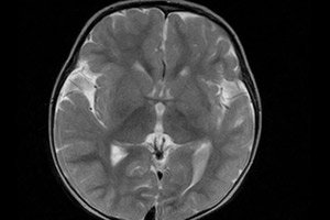
Today, the most informative instrumental diagnostics is considered to be using magnetic resonance imaging (MRI) of the brain. [ 13 ]
Electroencephalography is used to assess brain functions.
Differential diagnosis
Differential diagnosis is carried out with other congenital anomalies of the brain, including pachygyria, schizencephaly, syndromic disorders of cerebral functions, as well as idiopathic generalized and focal epilepsy in children. [ 14 ]
Who to contact?
Treatment polymicrogyria
In this congenital defect, treatment is aimed at eliminating symptoms. Thus, antiepileptic drugs are used to control seizures.
Other treatment methods: physiotherapy, occupational therapy, speech therapy.
Prevention
Given the significant proportion of spontaneous gene mutations leading to the development of this malformation of the cerebral cortex, prevention is considered impossible.
Forecast
In most cases, polymicrogyria has a poor prognosis: 87-94% of patients suffer from virtually incurable epilepsy with recurrent seizures. Many children with bilateral anomalies or lesions of more than half of the convolutions of one hemisphere die in early childhood.