
All iLive content is medically reviewed or fact checked to ensure as much factual accuracy as possible.
We have strict sourcing guidelines and only link to reputable media sites, academic research institutions and, whenever possible, medically peer reviewed studies. Note that the numbers in parentheses ([1], [2], etc.) are clickable links to these studies.
If you feel that any of our content is inaccurate, out-of-date, or otherwise questionable, please select it and press Ctrl + Enter.
The membranes of the brain
Medical expert of the article

Last reviewed: 04.07.2025

The brain, like the spinal cord, is surrounded by three meninges. These connective tissue sheets (meninges) cover the brain. The outermost of these meninges is the dura mater of the brain. Next to it is the middle one - the arachnoid, and inside of it is the inner soft (vascular) membrane of the brain, adjacent to the surface of the brain.
 [
[ Dura mater of the brain
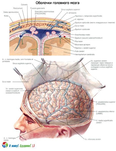
This membrane is distinguished by its special density, the presence of a large number of collagen and elastic fibers in its composition. The dura mater of the brain lines the cavities of the skull from the inside, and is also the periosteum of the inner surface of the bones of the brain section of the skull. The dura mater of the brain is loosely connected to the bones of the vault (roof) of the skull and is easily separated from them. In the area of the base of the skull, the membrane is firmly fused with the bones. The dura mater surrounds the cranial nerves emerging from the brain, forming their sheaths and merging with the edges of the openings through which these nerves leave the cranial cavity.
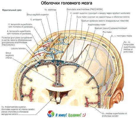
At the inner base of the skull (in the region of the medulla oblongata), the dura mater of the brain fuses with the edges of the foramen magnum and continues into the dura mater of the spinal cord. The inner surface of the dura mater, facing the brain (towards the arachnoid mater), is smooth, covered with flat cells. In some places, the dura mater of the brain is split. Its inner leaflet (duplicature) deeply intrudes in the form of processes into the cracks separating the parts of the brain from each other. In the places where the processes branch off (at their base), as well as in the areas where the dura mater attaches to the bones of the inner base of the skull, in the splits of the dura mater of the brain, triangular canals lined with endothelium are formed - the sinuses of the dura mater (sinus durae matris)
The largest process of the dura mater of the brain is the falx cerebri, or greater falx cerebri, located in the sagittal plane and penetrating the longitudinal fissure of the cerebrum between the right and left hemispheres. This is a thin, sickle-shaped curved plate of the dura mater, which penetrates the longitudinal fissure of the cerebrum in the form of two sheets. Without reaching the corpus callosum, this plate separates the right and left hemispheres of the cerebrum from each other. The superior sagittal sinus lies in the split base of the falx cerebri, which in its direction corresponds to the groove of the superior sagittal sinus of the cranial vault. The inferior sagittal sinus is located in the thickness of the free edge of the falx cerebri between its two sheets. In front, the falx cerebri is fused with the cock's crest of the ethmoid bone. The posterior part of the falx at the level of the internal occipital protrusion fuses with the tentorium cerebelli. Along the line of fusion of the posteroinferior edge of the falx cerebri and the tentorium cerebelli in the split of the dura mater of the brain there is a straight sinus, connecting the inferior sagittal sinus with the superior sagittal, transverse and occipital sinuses.
The tentorium cerebelli overhangs like a gable tent over the posterior cranial fossa, in which the cerebellum lies. Penetrating the transverse fissure, the tentorium cerebelli separates the occipital lobes of the cerebrum from the cerebellar hemispheres. The anterior edge of the tentorium cerebelli is uneven. It forms the incisura tentorii, to which the brainstem adjoins in front.
The lateral edges of the tentorium cerebelli are fused with the upper edge of the pyramids of the temporal bones. Behind, the tentorium cerebelli passes into the dura mater of the brain, lining the inside of the occipital bone. At the site of this transition, the dura mater of the brain forms a split - the transverse sinus, adjacent to the groove of the same name in the occipital bone.
The falx cerebelli, or lesser falx cerebelli, like the falx cerebri, is located in the sagittal plane. Its anterior edge is free and penetrates between the cerebellar hemispheres. The posterior edge (base) of the falx cerebelli continues to the right and left into the dura mater of the brain from the internal occipital protrusion above to the posterior edge of the foramen magnum below. The occipital sinus is formed at the base of the falx cerebelli.
Sella diaphragm
(diaphragma sellae) is a horizontally located plate with a hole in the center, stretched over the pituitary fossa and forming its roof. The pituitary gland is located under the diaphragm sellae in the fossa. Through the hole in the diaphragm, the pituitary gland is connected to the hypothalamus by means of a funnel.
Sinuses of the dura mater of the brain
The sinuses of the dura mater of the brain, formed by the splitting of the membrane into two plates, are channels through which venous blood flows from the brain into the internal jugular veins.
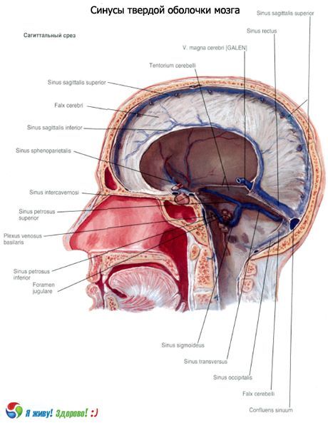
The sheets of the dura mater that form the sinus are tightly stretched and do not collapse. Therefore, the sinuses gape on section. The sinuses do not have valves. This structure of the sinuses allows venous blood to flow freely from the brain regardless of fluctuations in intracranial pressure. On the inner surfaces of the skull bones, in the locations of the sinuses of the dura mater, there are corresponding grooves. The following sinuses of the dura mater of the brain are distinguished.
- The superior sagittal sinus (sinus sagittalis superior) is located along the entire outer (upper) edge of the falx cerebri, from the cockscomb of the ethmoid bone to the internal occipital protuberance. In the anterior sections, this sinus has anastomoses with the veins of the nasal cavity. The posterior end of the sinus flows into the transverse sinus. To the right and left of the superior sagittal sinus are the lateral lacunae (lacunae laterales) communicating with it. These are small cavities between the outer and inner layers (sheets) of the dura mater of the brain, the number and size of which are very variable. The cavities of the lacunae communicate with the cavity of the superior sagittal sinus, and the veins of the dura mater of the brain, the veins of the brain, and the dyschiatic veins flow into them.
- The inferior sagittal sinus (sinus sagittalis inferior) is located in the thickness of the lower free edge of the falx cerebri. It is significantly smaller than the upper one. With its posterior end, the inferior sagittal sinus flows into the straight sinus, into its anterior part, in the place where the lower edge of the falx cerebri fuses with the anterior edge of the tentorium cerebelli
- The straight sinus (sinus rectus) is located sagittally in the cleft of the tentorium cerebelli along the line of attachment of the falx cerebri to it. The straight sinus connects the posterior ends of the superior and inferior sagittal sinuses. In addition to the inferior sagittal sinus, the great cerebral vein flows into the anterior end of the straight sinus. Behind, the straight sinus flows into the transverse sinus, into its middle part, which is called the sinus drain. The posterior part of the superior sagittal sinus and the occipital sinus also flow here.
- The transverse sinus (sinus transversus) is located at the point where the tentorium cerebelli departs from the dura mater of the brain. On the inner surface of the squama of the occipital bone, this sinus corresponds to a wide groove of the transverse sinus. The place where the superior sagittal, occipital and straight sinuses flow into it is called the sinus drain (confluens sinuum, confluence of sinuses). On the right and left, the transverse sinus continues into the sigmoid sinus of the corresponding side.
- The occipital sinus (sinus occipitalis) lies at the base of the falx cerebelli. Descending along the internal occipital crest, this sinus reaches the posterior edge of the foramen magnum, where it divides into two branches that encircle this opening from behind and from the sides. Each of the branches of the occipital sinus flows into the sigmoid sinus on its side, and the upper end into the transverse sinus.
- The sigmoid sinus (sinus sigmoideus) is paired, located in the groove of the same name on the inner surface of the skull, has an S-shape. In the area of the jugular foramen, the sigmoid sinus passes into the internal jugular vein.
- The cavernous sinus (sinus cavernosus) is paired and is located at the base of the skull on the side of the sella turcica. The internal carotid artery and some cranial nerves pass through this sinus. The sinus has a very complex structure in the form of caves communicating with each other, which is why it got its name. Between the right and left cavernous sinuses there are communications (anastomoses) in the form of the anterior and posterior intercavernous sinuses (sinus intercavernosi), which are located in the thickness of the diaphragm of the sella turcica, in front of and behind the pituitary infundibulum. The sphenoparietal sinus and the superior ophthalmic vein flow into the anterior sections of the cavernous sinus.
- The sphenoparietal sinus (sinus sphenoparietalis) is paired, adjacent to the free posterior edge of the lesser wing of the sphenoid bone, and is attached here by the dura mater of the brain in a split.
- The superior and inferior petrosal sinuses (sinus petrosus superior et sinus petrosus inferior) are paired and located along the superior and inferior edges of the pyramid of the temporal bone. Both sinuses participate in the formation of the outflow tracts of venous blood from the cavernous sinus to the sigmoid sinus. The right and left inferior petrosal sinuses are connected by several veins located in the cleft of the dura mater in the region of the body of the occipital bone, which are called the basilar plexus. This plexus is connected through the foramen magnum with the internal vertebral venous plexus.
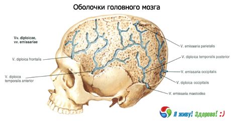
In some places, the sinuses of the dura mater of the brain form anastomoses with the external veins of the head with the help of emissary veins - graduates (vv. emissariae). In addition, the sinuses of the dura mater have communications with the diploic veins (vv. diploicae), located in the spongy substance of the bones of the cranial vault and flowing into the superficial veins of the head. Thus, venous blood from the brain flows through the systems of its superficial and deep veins into the sinuses of the dura mater of the brain and then into the right and left internal jugular veins.
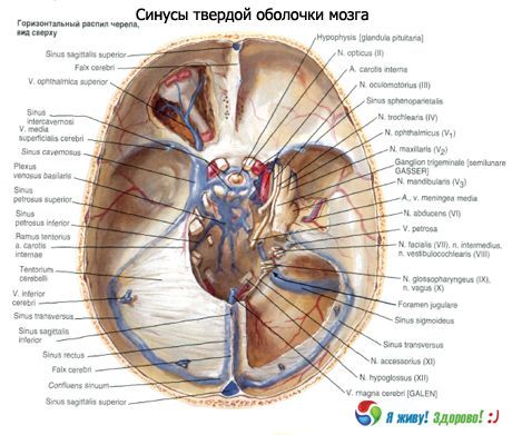
In addition, due to anastomoses of the sinuses with diploic veins, venous outlets and venous plexuses (vertebral, basilar, suboccipital, pterygoid, etc.), venous blood from the brain can flow into the superficial veins of the head and neck.
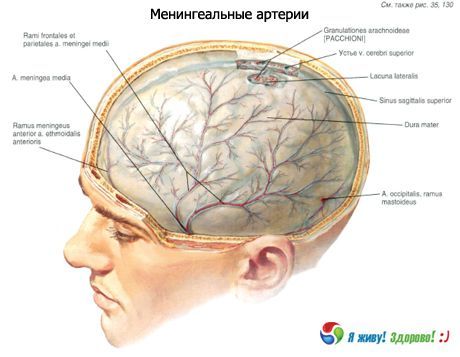
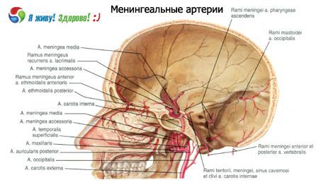
Vessels and nerves of the dura mater of the brain
The middle meningeal artery (a branch of the maxillary artery) approaches the dura mater of the brain through the right and left spinous openings and branches off in the temporoparietal region of the membrane. The dura mater of the brain, lining the anterior cranial fossa, is supplied with blood by branches of the anterior meningeal artery (a branch of the anterior ethmoidal artery from the ophthalmic artery). In the membrane of the posterior cranial fossa, the posterior meningeal artery branches off - a branch of the ascending pharyngeal artery from the external carotid artery, penetrating the cranial cavity through the jugular foramen, as well as the meningeal branches from the vertebral artery and the mammillary branch from the occipital artery, entering the cranial cavity through the mammillary foramen.
The veins of the pia mater of the brain flow into the nearest sinuses of the dura mater, as well as into the pterygoid venous plexus.
The dura mater of the brain is innervated by branches of the trigeminal and vagus nerves, as well as by sympathetic fibers entering the membrane in the thickness of the adventitia of the blood vessels. In the area of the anterior cranial fossa, it receives branches from the ophthalmic nerve (the first branch of the trigeminal nerve). A branch of this nerve, the tentorial (meningeal) branch, also supplies the tentorium cerebelli and the falx cerebri. The middle meningeal branch from the maxillary nerve, as well as a branch from the mandibular nerve (respectively the second and third branches of the trigeminal nerve), approach the membrane in the middle cerebral fossa.
Arachnoid mater of the brain
The arachnoid mater of the brain (arachnoidea mater encephali) is located medially from the dura mater of the brain. The thin, transparent arachnoid mater, unlike the pia mater (vascular), does not penetrate into the gaps between individual parts of the brain and into the grooves of the hemispheres. It covers the brain, passing from one part of the brain to another, and lies above the grooves. The arachnoid is separated from the pia mater of the brain by the subarachnoid space (cavitas subaracnoidalis), which contains cerebrospinal fluid. In places where the arachnoid mater is located above wide and deep grooves, the subarachnoid space is expanded and forms subarachnoid cisterns of greater or lesser size (cisternae subarachnoideae).
Above the convex parts of the brain and on the surface of the convolutions, the arachnoid and pia mater are tightly adjacent to each other. In such areas, the subarachnoid space narrows significantly, turning into a capillary gap.
The largest subarachnoid cisterns are the following.
- The cerebellomedullary cistern (cisterna cerebellomedullaris) is located in the depression between the medulla oblongata ventrally and the cerebellum dorsally. It is bounded posteriorly by the arachnoid mater. It is the largest of all the cisterns.
- The cistern of the lateral fossa of the cerebrum (cisterna fossae lateralis cerebri) is located on the lower lateral surface of the cerebral hemisphere in the fossa of the same name, which corresponds to the anterior parts of the lateral groove of the cerebral hemisphere.
- The cisterna chiasmatis is located at the base of the brain, anterior to the optic chiasm.
- The interpeduncular cistern (cisterna interpeduncularis) is located in the interpeduncular fossa between the cerebral peduncles, below (anteriorly) from the posterior perforated substance.
The subarachnoid space of the brain in the region of the foramen magnum communicates with the subarachnoid space of the spinal cord.
Cerebrospinal fluid
The cerebrospinal fluid (liquor cerebrospinalis), formed in the ventricles of the brain, is poor in protein substances and contains no cells. The total amount of this fluid is 100-200 ml. It is produced by the vascular plexuses of the lateral, III and IV ventricles from their blood capillaries. The walls of the blood capillaries, the basement membrane, the epithelial plate covering the capillaries form the so-called blood-brain barrier. This barrier of blood in the ventricular cavities selectively allows some substances to pass through and retains others, which is an important circumstance for protecting the brain from harmful effects.
From the lateral ventricles, through the right and left interventricular (Monroe's) openings, the cerebrospinal fluid enters the third ventricle, where there is also a choroid plexus. From the third ventricle, through the cerebral aqueduct, the cerebrospinal fluid enters the fourth ventricle and then through the unpaired opening in the posterior wall (Magendie's opening) and the paired lateral aperture (Lushka's opening), flows into the cerebellomedullary cistern of the subarachnoid space.
The arachnoid mater is connected to the soft mater lying on the surface of the brain by numerous thin bundles of collagen and elastic fibers, between which blood vessels pass. Near the sinuses of the dura mater of the brain, the arachnoid mater forms peculiar outgrowths, protrusions - granulations of the arachnoid mater (granulationes arachnoideae; Pachion's granulations). These protrusions protrude into the venous sinuses and lateral lacunae of the dura mater. On the inner surface of the bones of the skull, at the location of the granulations of the arachnoid mater, there are depressions - granulation pits, where the outflow of cerebrospinal fluid into the venous bed occurs.
Soft (vascular) membrane of the brain (pia mater encephali)
This is the innermost membrane of the brain. It is tightly attached to the outer surface of the brain and extends into all the crevices and furrows. The soft membrane consists of loose connective tissue, in the thickness of which are located blood vessels that go to the brain and feed it. In certain places, the soft membrane penetrates the cavities of the ventricles of the brain and forms vascular plexuses (plexus choroideus), which produce cerebrospinal fluid.
Age-related features of the membranes of the brain and spinal cord
The dura mater of the brain in a newborn is thin, tightly fused with the bones of the skull. The processes of the membrane are poorly developed. The sinuses of the dura mater of the brain are thin-walled, relatively wide. The length of the superior sagittal sinus in a newborn is 18-20 cm. The sinuses are projected differently than in an adult. For example, the sigmoid sinus is located 15 mm behind the tympanic ring of the external auditory canal. There is a greater asymmetry in the sizes of the sinuses than in an adult. The anterior end of the superior sagittal sinus anastomoses with the veins of the nasal mucosa. After 10 years, the structure and topography of the sinuses are the same as in an adult.
The arachnoid and pia mater of the brain and spinal cord in a newborn are thin and delicate. The subarachnoid space is relatively large. Its capacity is about 20 cm 3, increasing quite quickly: by the end of the 1st year of life up to 30 cm 3, by 5 years - up to 40-60 cm 3. In children aged 8 years, the volume of the subarachnoid space reaches 100-140 cm 3, in an adult it is 100-200 cm 3. The cerebellomedullary, interpeduncular and other cisterns at the base of the brain in a newborn are quite large. Thus, the height of the cerebellomedullary cistern is approximately 2 cm, and its width (at the upper border) is from 0.8 to 1.8 cm.
Использованная литература