
All iLive content is medically reviewed or fact checked to ensure as much factual accuracy as possible.
We have strict sourcing guidelines and only link to reputable media sites, academic research institutions and, whenever possible, medically peer reviewed studies. Note that the numbers in parentheses ([1], [2], etc.) are clickable links to these studies.
If you feel that any of our content is inaccurate, out-of-date, or otherwise questionable, please select it and press Ctrl + Enter.
Magnetic resonance imaging (MRI) of the brain
Medical expert of the article

Last reviewed: 03.07.2025
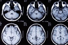
MRI of the brain is currently the leading non-invasive method of intravital visualization of the brain structure. Synonyms of MRI are nuclear magnetic resonance tomography and magnetic resonance imaging. The spatial resolution of the MRI method is 1-2 mm, it can be increased by contrasting with gadolinium.
 [
[ The purpose of performing MRI of the brain
The purpose of MRI of the brain is to identify and determine the shape, size and localization of various brain lesions [post-traumatic, atrophic, foci of ischemic (after 24 hours) and hemorrhagic (from the first hours) stroke, demyelinating processes, meningiomas and glial tumors], displacement of brain structures, severity of cerebral edema, the state of cerebrospinal fluid-containing spaces to exclude possible "organic" causes of psychopathological symptoms. MRI is also performed to diagnose brain and spinal lesions.
Indications for MRI of the brain
- Diagnosis of brain damage.
- Differential diagnostics of neuroinfections with non-infectious brain lesions.
- Monitoring the effectiveness of treatment of neuroinfections.
Indications for conducting research in a psychiatric clinic:
- suspicion of the presence of an atrophic, degenerative or demyelinating process, an epileptic focus, a stroke, a brain tumor.
Preparing for an MRI of the brain
Before MRI, the patient is informed about the procedure, its painlessness and absence of radiation, if a radioactive contrast agent is not used. In contrast MRI, the patient must be warned that after the introduction of the contrast agent, a feeling of heat and flushing, headache, metallic taste in the mouth, nausea or vomiting may occur.
The patient should be dressed in comfortable light clothing, all metal objects located in the field of the tomograph should be removed. In case of motor restlessness, anxiety, and claustrophobia, the patient is prescribed sedatives, as he must remain motionless during the examination.
The doctor must obtain written consent from the patient or his relatives to conduct the examination, and must also find out and note in the patient's medical history the presence of intolerance to iodine (seafood) and contrast agents. In case of allergic reactions to iodine, it is necessary to prescribe antihistamines prophylactically or cancel the administration of the contrast agent.
MRI brain research technique
The examination is carried out on a table, which is then moved into the cylindrical space of the scanner, in a supine position.
The doctor performing the examination changes the frequency of radio waves emitted by the scanner and adjusts the image quality using a computer.
Information about the sections is stored in digital form on a computer, displayed on a monitor and included in the medical record in the form of a photograph.
The method is based on the physical phenomenon of nuclear magnetic resonance. Nuclei of many atoms, in particular the nucleus of the hydrogen atom (proton), have a magnetic moment, which is associated with their rotation - spin. Such nuclei can be considered miniature elementary magnets. In a constant magnetic field, the spin can be located in the direction or against the magnetic lines of force, in these two cases the energy of the nucleus is different.
When exposed to an external radio-frequency pulsed magnetic field with certain parameters that cause magnetic resonance, the total magnetic field of the object, created by elementary magnets, changes and then decays to zero due to the reorientation of spins during the longitudinal relaxation time (Tj), as well as due to the disruption of the coherence of individual spins under the influence of the environment during the transverse relaxation time (T2).
These changes are registered by special sensors, and the magnitude of the received magnetic signal corresponds to the local concentration of nuclei, and the T1 and T2 values can be used to judge what chemical structures they are included in. Using computer processing, a picture of the distribution of the corresponding nuclei on the "sections" or in the volume of the brain is reproduced.
By using magnets that create high levels of magnetic field intensity, the signal can be subjected to spectral analysis, with the separation of components associated with not only hydrogen atoms, but also phosphorus (for example, to study the distribution of adenosine triphosphate metabolism), carbon, and fluorine. Since the exposure time (time resolution) is also reduced (to several seconds and even 100 ms), it is possible to study metabolic changes in various types of intellectual activity. This modification of the method, called "nuclear magnetic resonance spectroscopy" or "functional MRI", allows not only to visualize the structure, but also to study some functions of the brain.
Contraindications for MRI of the brain
- pregnancy;
- the presence of foreign metal and, especially, ferromagnetic objects on or in the patient's body, as well as electronic devices (in particular, watches, jewelry, metal staples on blood vessels, fragments), since exposure to a strong magnetic field can cause their displacement, heating or failure (for example, MRI is strictly contraindicated for patients with a wearable or implanted pacemaker).
Interpretation of MRI results
MRI evaluates the state of brain structures by their outlines, sizes and tissue density. It is important to note that MRI reflects tissue density depending on their water content, and therefore primarily identifies such lesions as cerebral edema-swelling (CED), demyelinating diseases, and tumors.
Since the highest concentration of protons is associated with water (intercellular fluid) and with lipids that form the myelin sheaths of nerve fibers, the MRI method clearly delineates the gray and white matter of the brain, visualizes spaces filled with fluid (ventricles of the brain, edema, cystic formations), allows for the diagnosis of atrophic and demyelinating processes, neoplasms, and also obtains volumetric distributions of a number of compounds (choline, lactate).
Factors influencing the result
A certain limitation of the MRI method (especially when using equipment that provides a relatively low magnetic field strength of 0.12-0.15 T) is the exposure duration, which can reach 10-15 minutes, when the patient must maintain a motionless posture (which is not always possible when examining children, the elderly and restless mentally ill patients). In these cases, muscle relaxants or anesthesia can be used [the use of anti-anxiety drugs (tranquilizers, anxiolytics) may be insufficient to relieve motor restlessness in patients], necessarily taking into account the ratio of the diagnostic information content of the study and the risk of possible complications from the use of drugs of the indicated groups.
Complications
The absence of ionizing radiation makes the MRI method highly safe, which determined its widespread use. Complications of the MRI method have not been described. Some improvement in cerebral blood flow in 10-15% of patients (which is associated with a change in the rheological properties of blood under the influence of a magnetic field) has been found as side effects.
When performing contrast MRI, the patient may have allergic reactions to the contrast agent in the form of a feeling of heat, headache, metallic taste in the mouth, nausea or vomiting. After completing a long study in a horizontal position, the patient may have orthostatic hypotension.
Alternative methods
In the absence of equipment for MRI, the best alternative is CT scanning, taking into account the features and limitations of the method.