
All iLive content is medically reviewed or fact checked to ensure as much factual accuracy as possible.
We have strict sourcing guidelines and only link to reputable media sites, academic research institutions and, whenever possible, medically peer reviewed studies. Note that the numbers in parentheses ([1], [2], etc.) are clickable links to these studies.
If you feel that any of our content is inaccurate, out-of-date, or otherwise questionable, please select it and press Ctrl + Enter.
The plague pathogen
Medical expert of the article

Last reviewed: 06.07.2025
Plague (pestis) is an acute infectious disease that occurs as a hemorrhagic septicemia. In the past, the plague was a terrible scourge for humanity. Three plague pandemics are known, which took millions of human lives.
The first pandemic occurred in the 6th century AD. It killed about 100 million people from 531 to 580 - half the population of the Eastern Roman Empire (the "Justinian" plague).
The second pandemic broke out in the 14th century. It began in China and affected many countries in Asia and Europe. In Asia, 40 million people died from it, and in Europe, out of 100 million people, 25 million died. This is how N. M. Karamzin describes this pandemic in his History of the Russian State: “The disease was revealed by glands in the soft cavities of the body, a person coughed up blood and died on the second or third day. It is impossible, the chroniclers say, to imagine a more terrible sight... From Beijing to the banks of the Euphrates and Ladoga, the bowels of the earth were filled with millions of corpses, and the states were deserted... In Glukhov and Belozersk, not a single inhabitant remained... This cruel plague came and returned several times. In Smolensk it raged three times, and finally, in 1387, only five people remained, who, according to the chronicle, went out and closed the city, which was filled with corpses.”
The third plague pandemic began in 1894 and ended in 1938, killing 13–15 million people.
The causative agent of the plague was discovered in 1894 by the French scientist A. Yersin, in whose honor it was named Yersinia pestis. The genus Yersinia belongs to the Enterobacteriaceae family and includes 11 species, of which three are pathogenic for humans: Yersinia pestis, Yersinia pseudotuberculosis and Yersinia enterocolitica; the pathogenicity of the others is still unclear.
Morphology of the plague pathogen
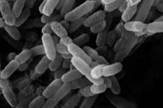
Yersinia pestis is 1-2 μm long and 0.3-0.7 μm thick. In smears from the patient's body and from the corpses of people and rodents who died from the plague, it looks like a short ovoid (egg-shaped) rod with bipolar staining. In smears from a broth culture, the rod is located in a chain, in smears from agar cultures - randomly. Bipolar staining is preserved in both cases, but is somewhat weaker in smears from agar cultures. The causative agent of the plague is Gram-negative, stains better with alkaline and carbolic dyes (Leffler's blue), does not form spores, and has no flagella. The G + C content in DNA is 45.8-46.0 mol % (for the entire genus). At a temperature of 37 ° C, it forms a delicate capsule of protein nature, which is revealed on moist and slightly acidic nutrient media.
Biochemical properties of the plague pathogen
Yersinia pestis is an aerobe, it grows well on regular nutrient media. The optimum temperature for growth is 27-28 °C (range - from 0 to 45 °C), pH = 6.9-7.1. The plague bacillus grows characteristically on liquid and solid nutrient media: on broth it manifests itself as the formation of a loose film, from which threads descend in the form of icicles, resembling stalactites, at the bottom - a loose sediment, the broth remains transparent. The development of colonies on solid media goes through three stages: after 10-12 hours under a microscope, growth in the form of colorless plates (the "broken glass" stage); after 18-24 hours - the "lace handkerchiefs" stage, when viewed under a microscope, a light lace zone is noticeable, located around the protruding central part, yellowish or slightly brownish in color. After 40-48 hours, the "adult colony" stage occurs - a brownish-outlined center with a distinct peripheral zone. Yersinia pseudotuberculosis and Yersinia enterocolitica do not have a "broken glass" stage. On media with blood, Yersinia pestis colonies are granular with a weakly defined peripheral zone. In order to quickly obtain growth characteristic of Yersinia pestis on media, it is recommended to add growth stimulants to them: sodium sulfite, blood (or its preparations) or sarcinia culture lysate. The plague bacillus is characterized by pronounced polymorphism, especially on media with an increased concentration of NaCl, in old cultures, in the organs of decomposed plague corpses.
The plague bacillus does not have oxidase, does not form indole and H2S, has catalase activity and ferments glucose, maltose, galactose, mannitol with the formation of acid without gas.
 [ 6 ], [ 7 ], [ 8 ], [ 9 ], [ 10 ]
Antigenic composition of the plague pathogen
Up to 18 similar somatic antigens have been found in Yersinia pestis, Yersinia pseudotuberculosis, and Yersinia enterocolitica. Yersinia pestis is characterized by the presence of a capsular antigen (fraction I), T, VW antigens, plasma coagulase proteins, fibrinolysin, outer membrane proteins, and pH6 antigen. However, unlike Yersinia pseudotuberculosis and Yersinia enterocolitica, Yersinia pestis is more uniform in antigen terms; there is no serological classification of this species.
Resistance of the plague pathogen
In sputum, the plague bacillus can survive for up to 10 days; on linen and clothing soiled with the patient's secretions, it survives for weeks (protein and mucus protect it from the destructive effect of drying). In the corpses of people and animals that died from the plague, it survives from early autumn until winter; low temperatures, freezing and thawing do not kill it. Sun, drying, and high temperatures are destructive for Yersinia pestis. Heating to 60 °C kills in 1 hour, at a temperature of 100 °C it dies in a few minutes; 70% alcohol, 5% phenol solution, 5% lysol solution and some other chemical disinfectants kill in 5-10-20 minutes.
Pathogenicity factors of the plague pathogen
Yersinia pestis is the most pathogenic and aggressive among bacteria, therefore it causes the most severe disease. In all animals sensitive to it and in humans, the plague pathogen suppresses the protective function of the phagocytic system. It penetrates phagocytes, suppresses the "oxidative burst" in them and reproduces unhindered. The inability of phagocytes to perform their killer function in relation to Yersinia pestis is the main reason for susceptibility to plague. High invasiveness, aggressiveness, toxigenicity, toxicity, allergenicity and the ability to suppress phagocytosis are due to the presence of a whole arsenal of pathogenicity factors in Y. pestis, which are listed below.
The ability of cells to absorb exogenous dyes and hemin. It is associated with the function of the iron transport system and provides Yersinia pestis with the ability to reproduce in the body's tissues.
- Dependence of growth at a temperature of 37 °C on the presence of Ca ions in the medium.
- Synthesis of VW antigens. Antigen W is located in the outer membrane, and V is in the cytoplasm. These antigens ensure the reproduction of Y. pestis inside macrophages.
- Synthesis of "mouse" toxin. The toxin blocks the process of electron transfer in the mitochondria of the heart and liver of sensitive animals, affects platelets and blood vessels (thrombocytopenia) and disrupts their functions.
- Synthesis of capsule (fraction I - Fral). The capsule inhibits the activity of macrophages.
- Pesticide synthesis is a species-specific characteristic of Yersinia pestis.
- Fibrinolysin synthesis.
- Synthesis of plasma coagulase. Both of these proteins are localized in the outer membrane and provide high invasive properties of Yersinia pestis.
- Synthesis of endogenous purines.
- Synthesis of heat-inducible proteins of the outer membrane - Yop proteins (Yersinia outer proteins). Proteins YopA, YopD, YopE, YopH, YopK, YopM, YopN suppress the activity of phagocytes.
- Synthesis of neuraminidase. It promotes adhesion (releases receptors for Yersinia pestis).
- Synthesis of adenylate cyclase. It is assumed that it suppresses the "oxidative burst", i.e. blocks the killing action of macrophages.
- Synthesis of adhesion pili. They inhibit phagocytosis and ensure the penetration of Yersinia pestis, as an intracellular parasite, into macrophages.
- Synthesis of broad-spectrum aminopeptidases.
- Endotoxin (LPS) and other cell wall components with toxic and allergenic effects.
- pHb-antigen. It is synthesized at a temperature of 37 °C and low pH, suppresses phagocytosis and has a cytotoxic effect on macrophages.
A significant part of the pathogenicity factors of Yersinia pestis is controlled by genes carried by the following 3 classes of plasmids, usually found together in all pathogenic strains:
- pYP (9.5 kb) - pathogenicity plasmid. Carries 3 genes:
- pst - encodes the synthesis of pesticin;
- pim - determines immunity to pesticide;
- pla - determines fibrinolytic (plasminogen activator) and plasma-coagulase activity.
- pYT (65 MD) is a toxigenicity plasmid. It carries genes that determine the synthesis of the "mouse" toxin (a complex protein consisting of two fragments, A and B, with m. w. 240 and 120 kDa, respectively), and genes that control the protein and lipoprotein components of the capsule. Its third component controls the chromosome genes. The plasmid was previously called pFra.
- pYV (110 kb) - virulence plasmid.
It determines the dependence of Y. pestis growth at 37 °C on the presence of Ca2+ ions in the medium, therefore it has another name - Lcr plasmid (low calcium response). The genes of this especially important plasmid also code for the synthesis of V and W antigens and heat-induced Yop proteins. Their synthesis is carried out under complex genetic control at a temperature of 37 °C and in the absence of Ca2+ in the medium. All types of Yop proteins, except YopM and YopN, are hydrolyzed due to the activity of the plasminogen activator (pla gene of the pYP plasmid). Yop proteins largely determine the virulence of Yersinia pestis. The YopE protein has antiphagocytic and cytotoxic effects. YopD ensures the penetration of YopE into the target cell; YopH has antiphagocytic and protein tyrosine phosphatase activity; the YopN protein has the properties of a calcium sensor; YopM binds to athrombin in human blood.
Post-infectious immunity
Post-infection immunity is strong and lifelong. Repeated plague cases are extremely rare. The nature of immunity is cellular. Although antibodies appear and play a certain role in acquired immunity, it is mediated mainly by T-lymphocytes and macrophages. In people who have had plague or been vaccinated, phagocytosis is complete. It is what determines acquired immunity.
Epidemiology of plague
The range of warm-blooded carriers of the plague microbe is extremely extensive and includes more than 200 species of 8 orders of mammals. The main source of plague in nature are rodents and lagomorphs. Natural infection has been established in more than 180 of their species, over 40 of which are part of the Fauna of Russia and adjacent territories (within the former USSR). Of the 60 species of fleas for which the possibility of transmitting the plague pathogen has been established under experimental conditions, 36 live in this territory.
The plague microbe multiplies in the lumen of the flea's digestive tract. In its anterior section, a plug is formed ("plague block"), containing a large number of microbes. When a mammal bites with a reverse blood flow into the wound, some of the microbes are washed off the plug, which leads to infection. In addition, excrement secreted by the flea during feeding can also cause infection if it gets into the wound.
The main (principal) carriers of Y. pestis in Russia and Central Asia are ground squirrels, gerbils and marmots, and in some foci also pikas and voles. The existence of the following plague foci is associated with them.
- 5 foci in which the main carrier of the plague microbe is the small ground squirrel (North-West Caspian region; Terek-Sunzha interfluve; Elbrus foci; Volga-Ural and Trans-Ural semi-desert foci).
- 5 foci in which the carriers are gophers and marmots (in Altai - pikas): Transbaikal, Gorno-Altai, Tuva and high-mountain Tien Shan and Pamir-Alai foci.
- Volga-Ural, Transcaucasian and Central Asian desert areas, where the main carriers are gerbils.
- High-mountain Transcaucasian and Gissar foci with the main carriers - voles.
Different classifications of Yersinia pestis are based on different groups of features - biochemical characteristics (glycerol-positive and glycerol-negative variants), area of distribution (oceanic and continental variants), types of main carriers (rat and ground squirrel variants). According to one of the most common classifications, proposed in 1951 by the French plague researcher R. Devignat, depending on the geographical distribution of the pathogen and its biochemical properties, three intraspecific forms (biovar) of Yersinia pestis are distinguished.
According to the classification of Russian scientists (Saratov, 1985), the species Yersinia pestis is divided into 5 subspecies: Yersinia pestis subsp. pestis (the main subspecies; it includes all three biovars of R. Devigny's classification), Y. pestis subsp. altaica (Altai subspecies), Yersinia pestis subsp. caucasica (Caucasian subspecies), Y. pestis subsp. hissarica (Gissar subspecies) and Yersinia pestis subsp. ulegeica (Udege subspecies).
Humans become infected through flea bites, direct contact with infectious material, airborne droplets, and rarely through food (for example, eating the meat of plague-infected camels). In 1998-1999, 30,534 people worldwide suffered from the plague, of whom 2,234 died.
Symptoms of the plague
Depending on the method of infection, there are bubonic, pulmonary, intestinal forms of plague; rarely, septic and cutaneous (purulent blisters at the site of a flea bite). The incubation period for plague varies from several hours to 9 days (in people undergoing seroprophylaxis, up to 12 days). The causative agent of plague penetrates through the smallest damage to the skin (flea bite), sometimes through the mucous membrane or by airborne droplets, reaches the regional lymph nodes, in which it begins to rapidly multiply. The disease begins suddenly: severe headache, high temperature with chills, the face is hyperemic, then it darkens, dark circles under the eyes ("black death"). A bubo (an enlarged inflamed lymph node) appears on the second day. Sometimes the plague develops so rapidly that the patient dies before the bubo appears. Pneumonic plague is especially severe. It can occur as a complication of bubonic plague, and through airborne infection. The disease also develops very rapidly: chills, high temperature, and already in the first hours pain in the side, cough, initially dry, and then with bloody sputum, are added; delirium, cyanosis, collapse, and death occurs. A patient with pneumonic plague is an exceptional danger to others, as he excretes a huge amount of the pathogen with sputum. In the development of the disease, the main role is played by the suppression of the activity of phagocytes: neutrophilic leukocytes and macrophages. Unrestrained reproduction and spread of the pathogen through the blood throughout the body completely suppresses the immune system and leads (in the absence of effective treatment) to the death of the patient.
Laboratory diagnostics of plague
Bacteriological, bacteriological, serological and biological methods are used, as well as an allergic test with pestin (for retrospective diagnostics). The material for the study is: a puncture from the bubo (or its discharge), sputum, blood, and, in the intestinal form, feces. Yersinia pestis is identified based on morphology, cultural, biochemical characteristics, a test with plague phage and using a biological test.
A simple and reliable method for determining plague bacillus antigens in the material being studied is the use of RPGA, especially with the use of erythrocyte diagnosticum sensitized with monoclonal antibodies to the capsular antigen, and IFM. These same reactions can be used to detect antibodies in the serum of patients.
The biological diagnostic method involves infecting a guinea pig with the test material (when it is heavily contaminated with accompanying microflora) cutaneously, subcutaneously, or, less commonly, intraperitoneally.
When working with material containing the plague pathogen, strict compliance with the regime is required, therefore all studies are carried out only by well-trained personnel in special anti-plague institutions.
Plague prevention
Constant monitoring of natural plague foci and organization of measures to prevent human diseases in the country is carried out by a special anti-plague service. It includes five anti-plague institutes and dozens of anti-plague stations and departments.
Despite the presence of natural foci, there has not been a single case of plague in humans in Russia since 1930. For specific plague prevention, a plague vaccination is used - a live attenuated vaccine from the EV strain. It is administered cutaneously, intradermally or subcutaneously. In addition, a dry tablet vaccine for oral use has been proposed. Post-vaccination immunity is formed by the 5th-6th day after vaccination and lasts for 11-12 months. An intradermal allergy test with pestin has been proposed for its assessment and retrospective diagnostics of plague. The reaction is considered positive if a seal of at least 10 mm in diameter is formed at the site of pestin administration after 24-48 hours and redness appears. The allergy test is also positive in people with post-infection immunity.
A great contribution to the study of the plague and the organization of the fight against it was made by Russian scientists: D. S. Samoylovich (the first not only in Russia, but also in Europe to “hunt” for the plague microbe back in the 18th century, he was also the first to propose vaccinations against the plague), D. K. Zabolotny, N. P. Klodnitsky, I. A. Deminsky (study of natural plague foci, carriers of the pathogen in the foci, etc.) and others.