
All iLive content is medically reviewed or fact checked to ensure as much factual accuracy as possible.
We have strict sourcing guidelines and only link to reputable media sites, academic research institutions and, whenever possible, medically peer reviewed studies. Note that the numbers in parentheses ([1], [2], etc.) are clickable links to these studies.
If you feel that any of our content is inaccurate, out-of-date, or otherwise questionable, please select it and press Ctrl + Enter.
Xeroderma pigmentosum
Medical expert of the article

Last reviewed: 04.07.2025
Xeroderma pigmentosum is an autosomal recessive genetic disorder of DNA repair. The disease is caused by mutations in genes that are involved in the repair of DNA damaged by ultraviolet radiation and other toxic substances.
Most often, this disease affects children, who are also called "children of the night." Frequent complications of the disease are basal cell carcinoma and other malignant neoplasms of the skin, metastatic malignant melanoma and squamous cell carcinoma.
 [ 1 ]
[ 1 ]
Causes xeroderma pigmentosum
Pathogenesis
Deficiency of UV endonuclease enzymes in the patient's cells (in fibroblasts) or their complete absence play an important role in the development of the disease. This enzyme is responsible for the reproduction of DNA damaged by ultraviolet rays. According to other sources, deficiency of DNA polymerase-1 enzyme plays an important role in the development of the disease in some patients. The disease most often develops under the influence of rays with a wavelength of 280-310 nm. It is believed that pigment xeroderma develops due to the entry of various photodynamic substances into the body or an increase in porphyrins in the human biological environment.
In the early stages of the disease, hyperkeratosis, atrophy of the epidermis focus, thinning of the Malpighian layer, an increase in melanin granules in the basal layer of cells, and chronic inflammation infiltrate in the upper layer of the dermis (mainly around blood vessels) are observed. Subsequently, degenerative changes are detected in collagen and elastic fibers, and at the tumor stage, signs characteristic of skin cancer are revealed.
Symptoms xeroderma pigmentosum
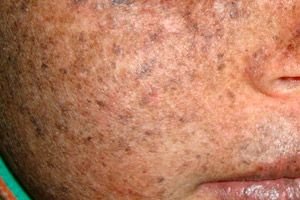
Men and women are equally affected by this disease. The disease begins in early childhood in spring or summer. The patient's skin is especially sensitive to sunlight. Therefore, the first rash in the form of hyperpigmentation, similar to a mole, appears on areas of the skin exposed to sunlight. The rash increases, erythema intensifies, becomes dark brown, telangiectasia, angioma and atrophy appear. Subsequently, papillomas and warts grow (mainly on the skin of the face and neck), spots and ulcers appear. As a result of ulcer scarring, the nose becomes thinner ("bird's beak"), the eyelid everts. Papillomas usually develop into malignant tumors. Pigmented xeroderma, as a rule, occurs together with spinocellular cancer, melanosarcomas.
What's bothering you?
Stages
In the clinical course of xeroderma pigmentosum, five stages are distinguished: inflammatory (erythematous), hyperpigmentation, atrophy, hyperkeratosis and malignant tumors.
During the inflammatory (erythematous) stage, swelling, red spots, and sometimes blisters and vesicles appear on areas of skin exposed to sunlight (face, neck, upper chest, arms, hands). There is almost no rash on areas not exposed to sunlight.
With hyperpigmentation, light brown, brown hyperpigmented spots similar to moles appear in place of the red spots.
With atrophy, the skin dries out, becomes thinner, and wrinkles. Numerous small, stellate telangiectasias and scars with a shiny surface are observed on the skin of the lips and nose. Due to atrophy and scars, microtomy (reduction of the mouth opening), ectropion, thinning of the ears and nose, atresia of the nasal opening and mouth develop. In 80-85% of patients, the eyes are affected: conjunctivitis, keratitis, damage to the cornea and mucous membrane, and weakening of vision are noted. Dyschromia, telangiectasia, hyperkeratotic changes and tumors are observed on the skin of the eyelids.
With hyperkeratosis, warty tumors, papilloma, keratoacanthoma, fibroma, angiofibrioma and other benign tumors appear in the above-described pathological foci. Therefore, some scientists include pigment xeroderma in the group of precancerous diseases.
After 10-15 years from the onset of the disease, malignant skin tumors (basalioma, endothelioma, angiosarcoma) appear in atrophically altered foci and pigment spots. The tumors are destroyed in a short time, metastasize to internal organs and lead to death. In the internal organs and tissues of some patients, general dystrophic changes are observed (syndactelia of the second and third toes, dental dystrophy, complete hair loss, etc.).
Forms
The neurological form of xeroderma pigmentosum manifests itself in two syndromes.
Reed syndrome is characterized by clinical manifestations of xeroderma pigmentosum and microcephaly, idiopathy, and slow growth of the skeletal system. In the neurological form of the disease, DNA reparation is difficult and X-ray therapy leads to an increase in the main pathological foci.
De Sinctis-X Cocchione syndrome is characterized by the following clinical signs:
- development of clinical signs of xeroderma pigmentosum on particularly photosensitive skin;
- early appearance of malignant tumors;
- simultaneous course of spastic paralysis, microcephaly and dementia;
- congenital deformities and dwarfism;
- underdevelopment of the gonads;
- frequent miscarriages;
- recessive transmission by inheritance.
According to some dermatologists, Santis-Cacchione syndrome is not an independent disease, but a severe and fully expressed clinical manifestation of xeroderma pigmentosum.
Genetic forms
Types |
Gene |
Locus |
Description |
Type A, I, XPA |
XPA |
9q22.3 |
Xeroderma pigmentosum (XP) group A – classic form |
Type B, II, XPB |
XPB |
2q21 |
XP Group B |
Type C, III, XPC |
XPC |
3p25 |
XP Group C |
Type D, IV, XPD |
XPD ERCC6 |
19q13.2-q13.3, 10q11 |
XP Group D or De Sanctis-Cacchione Syndrome |
Type E, V, XPE |
DDB2 |
11p12-p11 |
XP Group E |
Type F, VI, XPF |
ERCC4 |
16p13.3-p13.13 |
XP Group F |
Type G, VII, XPG |
RAD2ERCC5 |
13q33 |
XP group G and COFS syndrome (cerebro-oculo-facioskeletal syndrome) type 3 |
Type V, XPV |
POLH |
6p21.1-p12 |
Variant Xeroderma Pigmentosa |
What do need to examine?
How to examine?
Who to contact?
Treatment xeroderma pigmentosum
Antimalarial drugs (delagyl, plaquenil, resoquin, etc.), protecting DNA from ultraviolet rays, inhibit the depolymerization process, reduce the sensitivity (photosensitization) of the skin to sunlight. These drugs have anti-inflammatory and hyposensitizing properties. It is recommended to conduct general therapy together with vitamin therapy (B1, B2, PP, B6, B12, A, E), antihistamines (tavegil, diphenhydramine, suprastin), desensitizing agents (sodium thiosulfate, 10% calcium chloride intravenously 10 ml).
For local treatment, sunscreen creams and ointments are used.
In the tumor form of pigment xeroderma, surgical methods, liquid nitrogen and laser beams are used. To protect yourself from sunlight, you must wear loose clothing, sun hats and gloves.
Forecast
Most patients (2/3) die before reaching the age of 15. Some patients can live to 40-50 years.
[ 20 ]