
All iLive content is medically reviewed or fact checked to ensure as much factual accuracy as possible.
We have strict sourcing guidelines and only link to reputable media sites, academic research institutions and, whenever possible, medically peer reviewed studies. Note that the numbers in parentheses ([1], [2], etc.) are clickable links to these studies.
If you feel that any of our content is inaccurate, out-of-date, or otherwise questionable, please select it and press Ctrl + Enter.
eye socket
Medical expert of the article

Last reviewed: 06.07.2025
The orbit is a paired cavity resembling a four-sided pyramid with rounded edges. The base of the pyramid faces forward and forms the entrance to the orbit (aditus orbitae). The apex of the orbit is directed backward and medially. The optic canal (canalis opticus) passes through here. The eyeball, its muscles, lacrimal gland and other structures are located in the orbital cavity. The orbital cavity has four walls: superior, medial, inferior and lateral.
The superior wall is formed by the orbital part of the frontal bone and is only complemented by the lesser wing of the sphenoid bone at the back. At the border of the superior wall with the lateral wall of the orbit there is a shallow fossa of the lacrimal gland. At the medial edge of the superior wall, near the frontal notch, there is a barely noticeable depression - the trochlear fossa, next to which is located the trochlear spine.
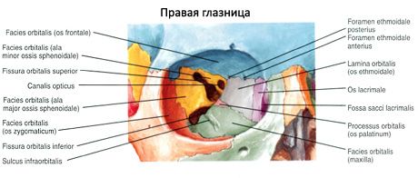
The medial wall is formed by the frontal process of the maxilla, the lacrimal bone, the orbital plate of the ethmoid bone, the body of the sphenoid bone (behind) and the medial part of the orbital part of the frontal bone (above). In the anterior part of the medial wall is the fossa of the lacrimal sac. Below, the fossa passes into the nasolacrimal canal (canalis nasolacrimal), which opens into the inferior nasal passage of the nasal cavity. Behind and above the fossa of the lacrimal sac, in the suture between the frontal bone and the orbital plate of the ethmoid bone, two openings are visible: the anterior ethmoidal opening (foramen ethmoidale anterius) and the posterior ethmoidal opening (foramen ethmoidale posterius) for the nerves and vessels of the same name.
The lower wall of the orbit is formed by the orbital surfaces of the maxilla and zygomatic bone. The wall is complemented at the back by the orbital process of the palatine bone. In the lower wall of the orbit is the infraorbital groove, which in front passes into the canal of the same name, opening on the anterior surface of the body of the maxilla as the infraorbital opening.
The lateral wall is formed by the orbital surfaces of the greater wing of the sphenoid bone and the frontal process of the zygomatic bone, as well as a small area of the zygomatic process of the frontal bone. Between the lateral and superior walls, deep in the orbit, is the superior orbital fissure, leading from the orbit into the middle cranial fossa. Between the lateral and inferior walls there is a large inferior orbital fissure (fissura orbitalis inferior); it is formed by the posterior margin of the orbital surface of the body of the maxilla, the orbital process of the palatine bone below, and the lower margin of the orbital surface of the greater wing of the sphenoid bone above. This fissure connects the orbit with the pterygopalatine and infratemporal fossae. On the lateral wall of the orbit there is a zygomaticoorbital foramen (for the zygomatic nerve), leading into a canal that, deep in the bone, divides into two canals. One of them opens on the lateral surface of the zygomatic bone through the zygomaticofacial foramen, the other - on the temporal surface through the zygomaticotemporal foramen.
What do need to examine?
How to examine?