
All iLive content is medically reviewed or fact checked to ensure as much factual accuracy as possible.
We have strict sourcing guidelines and only link to reputable media sites, academic research institutions and, whenever possible, medically peer reviewed studies. Note that the numbers in parentheses ([1], [2], etc.) are clickable links to these studies.
If you feel that any of our content is inaccurate, out-of-date, or otherwise questionable, please select it and press Ctrl + Enter.
Follicular conjunctivitis.
Medical expert of the article

Last reviewed: 12.07.2025
If the mucous membrane of the eye becomes inflamed with the appearance of vesicular formations - follicles (from the Latin folliculus - sac), then this is nothing more than follicular conjunctivitis. According to ICD-10, the code for the acute form of the disease is H10.019, and for the chronic form - H10.439.
Epidemiology
According to statistics, in 80% of cases, acute conjunctivitis, including follicular conjunctivitis, is caused by viruses, with adenoviruses accounting for 65-90% of cases.
The incidence of acute follicular conjunctivitis caused by HSV ranges from 1.3 to 4.8% of all cases of acute conjunctivitis.
Causes follicular conjunctivitis
Depending on the form of the inflammatory process, this type of conjunctivitis can be acute or chronic, and its types are determined by etiology.
Thus, the causes of acute follicular conjunctivitis include:
- respiratory adenoviruses of more than two dozen serotypes, causing adenoviral conjunctivitis and epidemic keratoconjunctivitis;
- HSV1 (herpes simplex virus) and Varicella zoster virus (herpes virus type 3 or chickenpox virus), infection with which leads to acute herpetic conjunctivitis. [ 1 ]
The main causes of chronic follicular conjunctivitis are recognized as:
- chlamydial infection – the bacterium Chlamydia trachomatis; [ 2 ]
- a viral skin infection – molluscum contagiosum, that is, a lesion of the skin of the eyelids, their edges and the mucous membrane of the eyes by a poxvirus (Molluscum contagiosum virus), which is transmitted by contact or through contaminated objects. [ 3 ]
Chronic inflammation of the conjunctiva may be associated with an allergy to locally applied ophthalmic drugs: eye drops (Proserin, Pilocarpine, Dipivefrin, [ 4 ] Carbachol, Atropine, Brinzolamide [ 5 ], etc.) or solutions of antiviral agents injected into the conjunctival sac.
The same infections also cause follicular conjunctivitis in children, more details in the publications:
Acute conjunctivitis in children
Risk factors
The most serious risk factor for developing infectious conjunctivitis is direct contact with exudate secreted from the patient's eyes or indirect contact, for example, through a towel or pillowcase on a pillow.
Common factors also include: poor personal hygiene; decreased immunity; the presence of ophthalmological diseases such as blepharitis, dry eye syndrome, inflammation of the meibomian glands of the eyelids or nasolacrimal duct; improper use of contact lenses, as well as prolonged use of certain eye drops.
Pathogenesis
In follicular conjunctivitis of viral origin, the pathogenesis is due to the fact that viral particles (virions) penetrate through the cytoplasmic membranes of epithelial cells into the cytoplasm and nuclei of cells. After the introduction of the viral nucleocapsid containing its genome (RNA or DNA), the structure of the cells of the mucous epithelium of the conjunctiva is disrupted, the virus begins to multiply: its DNA is transcribed and replicated in the cell nuclei.
In this case, some of the new virions are released from the nuclei and infect other cells, which leads to the activation of immunocompetent epithelial cells – T-lymphocytes, which destroy cells infected with the virus.
As studies have shown, the subconjunctival infiltrates in the form of follicles that form as a result of inflammation are accumulations of lymphocytes.
Symptoms follicular conjunctivitis
For most patients, the first signs of follicular conjunctivitis are redness of the eyes and a feeling of sand in the eyes.
When the conjunctiva is affected by adenovirus, the incubation period - from the moment of infection to the stage of the appearance of symptoms of inflammation - lasts approximately 10 days, and the duration of the disease can be 7-28 days.
The main symptoms are lacrimation and watery discharge (in case of chlamydial conjunctivitis – mucopurulent), swelling of the eyelids and diffuse swelling of the conjunctiva (chemosis), intolerance to bright light (photophobia), and blurred vision.
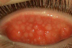
On the fornix of the conjunctiva (fornix conjunctivae) there appear pronounced bubble (papillary or vesicular) formations of a round shape, with a diameter of 0.5-1.5 mm.
Acute herpetic conjunctivitis, accompanied by itching and burning of the eyes, can have two forms: follicular and vesicular-ulcerative - with vesicular rashes on the eyelids (and serous discharge from them).
In the acute form, the lesion is often unilateral, but within a few days the second eye becomes infected. In almost half of the cases, there is an increase in the lymph nodes located in front of the ears and their pain during palpation - preauricular lymphadenopathy.
If the pharynx is inflamed at the same time (i.e. pharyngitis with sore throat occurs), an increase in body temperature is observed, which is defined as pharyngeal-conjunctival or pharyngoconjunctival fever.
Complications and consequences
A complication of herpetic keratoconjunctivitis is inflammation of the cornea of the eye and the development of herpetic keratitis.
A consequence of chronic follicular conjunctivitis caused by chlamydia can be trachoma - with inflammation of the superficial vessels of the cornea and its clouding.
Diagnostics follicular conjunctivitis
Follicular conjunctivitis is a clinical diagnosis and its diagnosis is made by careful examination of the eyes, examination of the conjunctiva and appropriate laboratory tests.
To determine the infection, the following tests are required: an eye smear (bacterial culture of secreted exudate) and a scraping from the conjunctiva, a general blood test, a blood test for antibodies to HSV1 and other viruses.
Differential diagnosis
Differential diagnostics are carried out with other types of conjunctivitis, as well as ophthalmological diseases that have similar symptoms (anterior uveitis, scleritis, etc.).
Who to contact?
Treatment follicular conjunctivitis
Treatment of follicular conjunctivitis caused by chlamydia includes not only topical agents but also oral antibacterial therapy using Tetracycline and Erythromycin.
Essential medications for topical use:
In case of inflammation of the mucous membrane of the eye caused by herpes viruses, ophthalmologists prescribe eye drops Trifluridine (Trifluridine, Lansurf, Viroptic) - one drop every two hours, and after three to four days - five times a day; eye gel Ganciclovir (Virgan) - up to five times during the day. Betadine (5% solution) is used - for lubricating the conjunctiva three times during the day.
Oral medications include Acyclovir (0.4 g three times daily), Valacyclovir ( Valtrovir ) (0.5 mg), or Famciclovir (0.25 g three times daily).
For the treatment of adenoviral conjunctivitis, antiviral treatment is not recommended; supportive measures to relieve symptoms include such agents as artificial tears or antihistamine drops (Cromogexal, Vizin, Opanadol, etc.), as well as cold compresses.
On the recommendation of a doctor, additional herbal treatment is possible, for more details see – Herbs for eye washing
In cases of molluscum contagiosum, surgical treatment can be performed - curettage, which is the mechanical removal of the affected layer of mucous tissue.
Prevention
In case of any conjunctivitis of infectious origin, preventive measures include observing the rules of personal hygiene, first of all, hand cleanliness.
Hands should always be washed with soap, and if there has been contact with a person who has conjunctivitis, hands should be treated with an alcohol-based disinfectant.
Forecast
With follicular conjunctivitis, the prognosis for the vast majority of patients is favorable.