
All iLive content is medically reviewed or fact checked to ensure as much factual accuracy as possible.
We have strict sourcing guidelines and only link to reputable media sites, academic research institutions and, whenever possible, medically peer reviewed studies. Note that the numbers in parentheses ([1], [2], etc.) are clickable links to these studies.
If you feel that any of our content is inaccurate, out-of-date, or otherwise questionable, please select it and press Ctrl + Enter.
Study will help develop therapy to improve lung function in fetuses with growth retardation
Last reviewed: 02.07.2025

If the fetus grows below normal levels during pregnancy, the risk increases with each week of gestation that some of its organs may not develop properly, which can negatively affect the baby's health after birth. The long-term effects of fetal growth restriction on brain and cardiovascular development have been the subject of much research, but there is a lack of scientific data on its effects on the lungs.
This was the subject of a study conducted jointly by the BCNatal Fetal Medicine Research Centre (Clínic Barcelona and Sant Joan de Déu Hospitals) and the Pompeu Fabra University (UPF), which identified differences in lung development between fetuses with limited growth and normal fetuses in terms of their vascular resistance. The researchers studied this by measuring the blood flow velocity in the fetus and analysing this data with the support of artificial intelligence methods and computer models.
The findings, recently published in a paper in the journal Scientific Reports, open the possibility of developing therapies aimed at improving lung development in growth-restricted fetuses and preventing respiratory problems that can persist not only into childhood but also into adolescence and adulthood.
The lead investigators of this study are Fátima Crispiel, a BCNatal and Clínic-IDIBAPS researcher in the Fetal and Perinatal Medicine Group, and Bart Beijnens (ICREA, UPF), a researcher in the BCN MedTech Unit in the UPF Engineering Department. The other researchers belong to different services and research groups of Clínic-IDIBAPS and are also associated with the University of Barcelona and the CIBER for Respiratory and Rare Diseases.
The study involved more than 200 pregnant women. The study analyzed fetal blood flow and its changes with additional oxygen in 208 pregnant women between 24 and 37 weeks of pregnancy. All women were examined at the Clínic Hospital in Barcelona, where they underwent all the necessary tests for the study.
In 97 of these cases, the fetuses had limited growth retardation, resulting in very low birth weight. The remaining 111 fetuses had normal growth. In each of these fetuses, the blood flow velocity in the main arteries and pulmonary vessels was measured, then compared using artificial intelligence. In addition, the resistance of the lungs was calculated using a computer model.
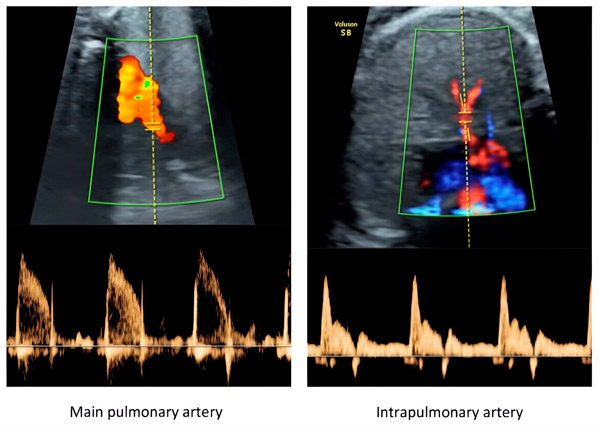
Illustrative Doppler images of the fetal main pulmonary artery and intrapulmonary artery. Source: Vellvé, K., Garcia-Canadilla, P., Nogueira, M., et al.
Fetal lung blood flow velocity was analyzed both under normal maternal breathing conditions and after supplemental oxygen was administered via a mask (hyperoxygenation conditions). This analysis was performed using a technique based on the emission of ultrasound waves to the fetus to estimate the blood flow velocity throughout its circulation based on Doppler principles.
In contrast, the resistance of organs such as the lungs cannot be measured directly using ultrasound, and a computer model representing the heart and blood vessels was used to measure it. For comparison, this computer model can be compared to a simulation of an electronic circuit. The researchers recreated a computer version of the fetal vascular system and, using the measured blood flow rates and modeling other parameters, were able to estimate the resistance and elasticity of the various organs.
Finally, machine learning methods based on artificial intelligence techniques were used to compare the blood flow patterns of the fetuses, which helped to group them into different categories according to flow parameters and clinical indicators.
Subsequently, examination of the effects of hyperoxygenation showed that it was associated with changes in lung resistance as a result of the extra oxygen supplied to the mother, and more oxygen was shown to improve pulmonary blood flow in growth-restricted fetuses without affecting normal fetuses.
"Basically, the results of the study show that growth-restricted fetuses have a different mean blood flow velocity as well as vascular resistance in the lungs than normal fetuses, and this can be normalized by providing the mother with additional oxygen," explains Beijnens (ICREA, UPF).
"The discovery of these differences in the pulmonary vessels opens the possibility of developing future therapeutic strategies to improve lung function in fetuses with growth restriction. After birth, these improvements in fetal development may reduce the risk of developing respiratory diseases later in life," explains Dr. Crispius (BCNatal, Clínic).