
All iLive content is medically reviewed or fact checked to ensure as much factual accuracy as possible.
We have strict sourcing guidelines and only link to reputable media sites, academic research institutions and, whenever possible, medically peer reviewed studies. Note that the numbers in parentheses ([1], [2], etc.) are clickable links to these studies.
If you feel that any of our content is inaccurate, out-of-date, or otherwise questionable, please select it and press Ctrl + Enter.
Orbital tumors in children
Medical expert of the article

Last reviewed: 07.07.2025
Various vascular tumors can be localized in the orbit. The following neoplasms are most common in children.
Capillary hemangioma
The most common orbital tumor occurring in childhood. It occurs more often in girls than in boys. A characteristic feature is the possibility of spontaneous regression. Clinical symptoms of capillary hemangioma:
- most often localized on the upper eyelid or in the orbit;
- During the first months of a child's life, the tumor is characterized by rapid growth, followed by a period of slow regression;
- exophthalmos;
- Amblyopia is usually caused by anisometropia, and sometimes by strabismus or deprivation (as a result of severe ptosis).
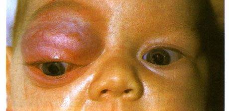
Capillary hemangioma of the anterior orbit and upper eyelid. The neoplasm has a tendency to progress
Since spontaneous regression is observed in almost all cases, treatment is prescribed only if there is a risk of developing amblyopia.
 [ 1 ], [ 2 ], [ 3 ], [ 4 ], [ 5 ], [ 6 ], [ 7 ]
Hemangiopericytoma
A rare tumor originating from adventitial cells (Rouget cells). It usually occurs in adults. It is characterized by invasive growth and is capable of forming distant metastases. The most typical clinical manifestation is increasing exophthalmos.
Lymphohemangioma
A vascular neoplasm, often developing in childhood. Differential diagnosis with hemangioma is difficult. However, unlike capillary hemangioma, the tumor is not prone to either progression or spontaneous regression. The neoplasm may be located superficially, or it may be localized deep in the orbit, manifesting as exophthalmos. As long as the functions remain high, surgical intervention is not indicated.
Congenital varicose veins of the orbit
Without contrast vasography of the orbit, it is difficult to differentiate this pathology from lymphangioma. Varicose veins manifest themselves as recurrent hemorrhages and suddenly appearing, rapidly increasing exophthalmos. In mild cases of exophthalmos, conservative treatment is limited.
What do need to examine?
How to examine?