
All iLive content is medically reviewed or fact checked to ensure as much factual accuracy as possible.
We have strict sourcing guidelines and only link to reputable media sites, academic research institutions and, whenever possible, medically peer reviewed studies. Note that the numbers in parentheses ([1], [2], etc.) are clickable links to these studies.
If you feel that any of our content is inaccurate, out-of-date, or otherwise questionable, please select it and press Ctrl + Enter.
Tumor cells: what they are, properties, features
Medical expert of the article

Last reviewed: 04.07.2025
Today, many people wonder what tumor cells are, what their role is, whether they are dangerous or beneficial, or are they solely aimed at destroying the macroorganism? Let's look into this issue.
Transformed cells that form a malignant tumor. The cells undergo numerous changes. These changes are noticeable at the morphological, chemical, and biochemical levels. Some are visible even to the naked eye. Detection of others requires special equipment. Everything depends on the type and location.
A distinctive feature is the ability to increase its biomass indefinitely, which is caused by a violation of apoptosis (provides programmed death). Such growth ends only with the death of a person.
The difference between a tumor cell and a normal cell
There is a system of cellular apoptosis, which is a programmed death of a cellular link. Usually, a cell that has completed its life cycle dies. In its place, a new subpopulation of the cell cycle develops over time. But during cancer transformation, such a natural mechanism is disrupted, as a result of which this cell does not die, but continues to grow and function in the body.
It is this internal mechanism that is the basic foundation of tumor formation, which has a tendency to uncontrolled and unlimited growth. That is, in essence, this kind of cellular structure is a cell that is incapable of death and has unlimited growth.
 [ 1 ], [ 2 ], [ 3 ], [ 4 ], [ 5 ]
Cellular atypia and atypical cells
Atypical cells are cells subject to mutation. Most often, atypical cells are formed under the influence of various external factors or heredity through their transformation from stem cells. Most often, the trigger for the development of a tumor cell is a specific gene that codes for cell death. Some potentially oncogenic viruses, such as retroviruses and herpes viruses, are capable of causing the transformation of stem cells into cancer cells.
Cellular atypism is the actual process of transformation that healthy cells undergo. This process includes a complex of chemical and biochemical processes. Mutation occurs under conditions of immune system disorders, especially in autoimmune diseases, in which the function of the immune system is transformed in such a way that it begins to produce antibodies directed against the cells and tissues of the body itself. The development of cellular atypism is facilitated by the deterioration of the body's natural defenses, in particular, with a violation of the activity of T-lymphocytes (killers), the processes of cell death are disrupted, which leads to their malignant degeneration.
[ 6 ], [ 7 ], [ 8 ], [ 9 ], [ 10 ], [ 11 ]
Carcinogenesis
The process of potential tissue growth, which is in no way associated with the normal state of the body. Carcinogenesis implies the process of degeneration of a normal cell into a tumor cell, which is a local formation, but the entire body is involved. Characteristic - tumors can metastasize, grow endlessly.
[ 12 ], [ 13 ], [ 14 ], [ 15 ], [ 16 ], [ 17 ], [ 18 ]
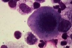
Cancer cell under a microscope
The development of a cancer cell is based on a sharp increase in the nucleus. A cancer cell is easy to detect under a microscope, since the nucleus can occupy most of the cytoplasm. The mitotic apparatus is also clearly expressed, and its violations are noticeable. First of all, the presence of chromosomal aberrations and non-disjunction of chromosomes attracts attention. This leads to the formation of multinuclear cells, an increase and thickening of the nucleus, and their transition to the mitotic division phase.
Deep invaginations of the nuclear membrane can also be detected under a microscope. Electron microscopy reveals intranuclear structures (granules). Light microscopy can also reveal loss of clarity of the nuclear contours. Nucleoli can retain normal configuration, and can increase in quantity and quality.
Swelling of mitochondria occurs. At the same time, the number of mitochondria decreases, mitochondrial structures are disrupted. Also, a diffuse arrangement of ribosomes relative to the endoplasmic reticulum is observed. In some cases, the Golgi apparatus may completely disappear, but in some cases, its hypertrophy is also possible. Subcellular structures also change, for example, the structure and appearance of lysosomes and ribosomes change. In this case, unequal degrees of differentiation of cellular structures occur.
Microscopy can reveal low-differentiated and highly differentiated tumors. Low-differentiated tumors are pale cells that contain a minimum number of organelles. The cell nucleus occupies most of the cellular space. At the same time, all subcellular structures have different degrees of maturity and differentiation. Highly differentiated tumors are characterized by the preservation of the original tissue structure.
Properties and characteristics of tumor cells
If a cell becomes tumorous, its genetic structure is disrupted. This entails repression processes. As a result of derepression of other genes, modified proteins, isoenzymes appear, and cell division occurs. This can change the intensity of gene and enzyme functioning. Repression of protein components is often observed. Previously, they were responsible for cell specialization, and were activated by depression.
Tumor transformation of a cell
Elements that act as triggers that initiate the pathological process. There is an assumption that the introduction of chemicals is carried out directly into the DNA and RNA of cells. This contributes to the disruption of maturation, an increase in cellular permeability develops, as a result of which potentially oncogenic viruses are able to penetrate the cell.
Some physical factors, such as increased radiation levels, irradiation, and mechanical factors, can also act as triggers. As a result of their impact, damage to the genetic apparatus, disruption of the cell cycle, and mutations occur.
Consumption of amino acids increases sharply, anabolism increases, while catabolic processes decrease. Glycolysis increases sharply. There is also a sharp decrease in the number of respiratory enzymes. A change in the antigen structure of the tumor cell is also observed. In particular, it begins to produce alpha-fetoprotein protein.
Markers
The simplest way to diagnose an oncological disease is to take a blood test to detect tumor markers. The test is carried out quite quickly: 2-3 days, in case of emergency it can be done in 3-4 hours. During the analysis, specific markers are identified that indicate the occurrence of oncological processes in the body. By the type of marker identified, it is possible to talk about what type of cancer is occurring in the body, and even determine its stage.
Atypism
It should be understood that the cell is not capable of death. It can also give pathological metastases. It is also characterized by a violation of synthetic processes, intensively absorbs glucose, quickly breaks down proteins and carbohydrates, changes the action of enzymes.
Genome
The very essence of transformational changes is the activation of nucleic acid synthesis. The standard complex undergoes significant changes. The synthesis of DNA polymerase-3, which is responsible for the synthesis of new DNA based on the native structure, is reduced. Instead, the synthesis of similar structures of type 2 increases, which is capable of restoring DNA even based on denatured DNA. This is what provides the specificity of the elements under consideration.
Receptors
The most well-known is the epidermal growth factor receptor, which is a transmembrane receptor. It actively interacts with epidermal growth factors.
Immunophenotype
Any transformation entails a change in the genotype. This is clearly expressed in changes that are reflected at the phenotypic level. Any change of this kind is foreign to the organism. This implies excessive aggressiveness of the human immune system, which is accompanied by an attack and destruction of the organism's own tissues.
Tumor cell expression
Expression is explained by several reasons. Only one cell is involved in primary carcinogenesis, but sometimes several cells can be involved in this process at the same time. Then a tumor develops, grows and multiplies. Often the process is accompanied by spontaneous mutations. Tumors acquire new properties.
A distinctive feature is the ability to express genes that act as growth factors for the tumor. They completely change the metabolic processes of the original cell, subordinating it to their needs, acting as a kind of parasite.
[ 24 ], [ 25 ], [ 26 ], [ 27 ], [ 28 ], [ 29 ], [ 30 ], [ 31 ]
Diffuse expression
For active cell division, the presence in the blood of a constant expression of a factor that suppresses (represses) gene activity is required.
[ 32 ], [ 33 ], [ 34 ], [ 35 ], [ 36 ], [ 37 ], [ 38 ], [ 39 ]
Lack of expression
During differentiation of mutated tissue, it loses the ability to express the reducing gene, which is responsible for programmed apoptosis. The loss of this ability deprives the corresponding structure of the ability to cease to exist. Accordingly, it continuously grows and multiplies.
[ 40 ], [ 41 ], [ 42 ], [ 43 ], [ 44 ]
Proliferation of tumor cells
Proliferation is an indicator of growth, determines the severity and stage. Functional anaplasia is observed. Rapidly growing tumors completely lose all the original properties of the tissue.
Proliferation index
The indicator depends on the localization. It is determined by the expression of Ki-67. It is expressed as a percentage by determining the ratio between the number of normal cells and the number of tumor cells. It is expressed as a percentage, where 1% is the minimum number, the early stage of the tumor process. 100% is the maximum stage, usually detected in a fatal outcome.
Uniqueness
They are transformed cells that have undergone mutation processes. These cells also have a pronounced ability to transform the basic properties of the original cell. A distinctive feature is the inability to die and the ability to grow without limit.
Uniformity
First of all, it is necessary to know that this phenomenon is nothing more than a degenerated cell of the human body, which for various reasons has undergone a malignant transformation. Almost any healthy cell of the human body can potentially undergo this process. The main thing is the presence of a trigger factor that will launch the mechanism of transformation (carcinogenesis). Such factors can be a virus, damage to the cellular or tissue structure, the presence of a special gene encoding cancerous degeneration.
Circulating tumor cells
The main feature of such a cell is a change in its biochemical cycle. There is a change in enzymatic activity. It is also worth noting the tendency to reduce the amount of DNA polymerase 3, which uses all components of the native DNA of the cell. Synthesis also changes significantly. Protein synthesis increases sharply, both qualitatively and quantitatively. Of particular interest is the presence of a large-nuclear spindle protein in cancer cells. Normally, the content of this protein should not exceed 11%, with tumors, the number increases to 30%. Metabolic activity changes.
[ 45 ], [ 46 ], [ 47 ], [ 48 ], [ 49 ]
Tumor stem cells
It can be said that these are primary, undifferentiated structures that will subsequently undergo differentiation of functions. If such a cell undergoes mutation and turns into a cancer cell, it becomes a source of metastases, since it moves freely with the blood flow and is capable of differentiating into any tissue. It lives a long time and proliferates slowly. When transplanted to someone with low immunity (immunodeficiency), it can cause the development of a malignant neoplasm
Apoptosis of tumor cells
The main problem of a tumor cell is that it has disrupted apoptosis processes (programmed death, it is not capable of death, and continues to grow and multiply constantly). There is a gene that inactivates the gene that makes the cell immortal. This allows you to restart the apoptosis processes, as a result of which you can establish normal cellular processes and return the cell to a normal state, causing its death.
[ 50 ], [ 51 ], [ 52 ], [ 53 ], [ 54 ], [ 55 ], [ 56 ], [ 57 ]
Differentiation of tumor cells
Tumor cells are differentiated depending on the tissues they are part of. The names of tumors also depend on the names of the tissues they are part of, as well as on the organ that has undergone tumor transformation: myoma, fibromyoma, epithelial, connective tissue tumor.
Использованная литература