
All iLive content is medically reviewed or fact checked to ensure as much factual accuracy as possible.
We have strict sourcing guidelines and only link to reputable media sites, academic research institutions and, whenever possible, medically peer reviewed studies. Note that the numbers in parentheses ([1], [2], etc.) are clickable links to these studies.
If you feel that any of our content is inaccurate, out-of-date, or otherwise questionable, please select it and press Ctrl + Enter.
Tuberous sclerosis
Medical expert of the article

Last reviewed: 04.07.2025
Tuberous sclerosis (synonyms: Pripgl-Burnewelli disease, Burnewelli-van der Heve phakomatosis, etc.) is a hereditary disease characterized by hyperplasia of ecto- and mesoderm derivatives. The inheritance type is autosomal dominant. Mutant genes are located in loci 16p13 and 9q34 and encode tuberins - proteins that regulate the GT-phase activity of other extracellular proteins.
Causes tuberous sclerosis
Tuberous sclerosis is a multisystem disease affecting the ectoderm derivatives (skin, nervous system, retina) and mesoderm (kidneys, heart, lungs). Inheritance is autosomal dominant with variable expressivity and incomplete penetrance. Linkage to the llql4-1 lq23 locus has been established. The presence of other defective genes is likely, in particular, those located on chromosomes 12 and 16. Up to 86% of cases of the disease are a consequence of new mutations, however, with a thorough comprehensive examination of relatives of patients, including skull tomography, eye and kidney examination, the number of hereditary forms increases.
Pathogenesis
In angiofibromas, fibroblast proliferation, collagen fiber growth, new vessel formation, capillary dilation, and absence of elastic fibers are observed. In hypopigmented spots, a decrease in melanocytes and melanosome size, and a decreased melanin content in melanocytes and keratinocytes are observed.
Pathomorphology
Angiofibromas consist of a large number of small vessels, often with widened lumens, located in dense connective tissue with a large number of cellular elements. Over time, the sebaceous glands become atrophic and even disappear completely. The number of hair follicles, however, is often increased, sometimes they are immature. Soft fibromas histologically have a typical picture of fibroma, but without a vascular component. In the area of shagreen foci, in the lower part of the reticular layer of the dermis, massive proliferations of homogeneous collagen fibers are visible, which resembles scleroderma. Elastic fibers in these places are fragmented, vessels and skin appendages are absent. Ultrastructural examination reveals dense compact bundles of collagen fibrils, among which there are single curved or twisted fibrils located among a fine-fibrous substance, possibly a precursor of collagen. In the foci of hypopigmented spots, although a normal number of melanocytes is noted, however, they, as well as the epithelial cells, do not contain pigment. In the melanocytes of white spots, an electron microscopic study revealed a decrease in the size of melanosomes and a decrease in the content of mature melanin. In milky white spots, melanosomes are found only in the initial stages of development, as in cutaneous-ocular albinism.
The histogenesis is still unclear, but an increase in the sensitivity of lymphocytes and fibroblasts of patients with tuberous sclerosis to ionizing radiation has been found, which may indicate a disruption of DNA repair processes and explain the frequency of new mutations.
Symptoms tuberous sclerosis
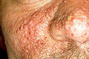
The disease begins in childhood or adolescence. The skin is affected in 96% of cases. Characteristic are the presence of nodules the size of a pinhead to a pea, located symmetrically in the nasolabial folds, on the chin, in the parotid region. The nodules are round, oval, flattened, brownish-red in color, usually closely adjacent to each other, sometimes merge, and protrude above the surrounding skin. Their surface is smooth, often with telangiectasias. On the body, "shagreen" plaques are noted, which are a connective tissue nevus. They are slightly raised above the skin surface, soft, with a bumpy surface resembling an orange peel. There are periungual fibromas (Koenen's tumor) - tumors or nodes on the nail fold. 80% of patients have hypopigmented white spots with a yellowish or grayish tint, located on the trunk, legs, arms and neck.
Clinically, the classic triad of symptoms is characteristic: angiofibromas, mental retardation and epilepsy. Symmetrical distribution of angiofibromas on the face is noted, mainly in the nasolabial folds, on the cheeks, chin, less often on the forehead and scalp. They are small reddish nodules with a smooth surface, usually appearing in childhood, and are found in 90% of patients over the age of 4. In addition to angiofibromas, fibromas, shagreen-like lesions, "café au lait" spots, hypopigmented spots, subungual and periungual fibromas and nodules on the oral mucosa are found on the skin.
Soft, pale pink or brown, tumor-like or plaque-like fibromas of various sizes are usually localized on the forehead, scalp, and upper cheeks. Shagreen-like lesions, along with angiofibromas, are the most common skin manifestations of tuberous sclerosis. They are detected in almost all patients over 5 years of age as flat, raised lesions of various sizes, the color of normal skin and a "lemon peel" surface. They are usually located in the lumbosacral region. "Coffee-with-milk" spots, as shown by the studies of SD Bell and DM MacDonald (1985), are equally common in patients with tuberous sclerosis and in healthy individuals, and therefore have no diagnostic value. In contrast, hypopigmented spots are important for diagnosis. They usually resemble the outline of a leaf, pointed on one side and rounded on the other, and have a pale grayish or milky white color. In people with light skin, the spots can only be seen with a Wood's lamp. They exist from birth and only increase in size with age. The combination of spots with epileptiform seizures in children has diagnostic value. Subungual and periungual tumor-like formations are fibromas or angiofibromas. Retinal tumors - phacomas, or astrocytic hamartomas - are also one of the most common manifestations of tuberous sclerosis. Although they are nonspecific, they are characteristic of tuberous sclerosis, so an eye examination is necessary in all cases if tuberous sclerosis is suspected. Another characteristic feature is intracranial calcifications, which are revealed by X-ray examination, which gave the disease the name "tuberous sclerosis". Convulsions are often the earliest symptoms of the disease and are considered epilepsy until skin symptoms appear. Less common manifestations of tuberous sclerosis are skeletal abnormalities, rhabdomyomas, tumors of the nervous system, visceral organs, and dysembryoplasia.
The full clinical picture of the disease also includes intracerebral calcifications, retinal tumors, hemarthromas and cysts of the kidneys, hemarthromas of the liver, and cardiac rhabdomyoma.
What do need to examine?
How to examine?
Who to contact?
Treatment tuberous sclerosis
The nodules are removed using a laser or electrocoagulation.
Forecast
The prognosis depends on changes in the brain and internal organs.
 [ 16 ]