
All iLive content is medically reviewed or fact checked to ensure as much factual accuracy as possible.
We have strict sourcing guidelines and only link to reputable media sites, academic research institutions and, whenever possible, medically peer reviewed studies. Note that the numbers in parentheses ([1], [2], etc.) are clickable links to these studies.
If you feel that any of our content is inaccurate, out-of-date, or otherwise questionable, please select it and press Ctrl + Enter.
Pharyngeal examination
Medical expert of the article

Last reviewed: 07.07.2025
Examination of the pharynx includes a number of procedures aimed at identifying both local and manifestations of general diseases, as well as pathological conditions caused by impaired innervation of the organs of the oral cavity and pharynx. If neurological disorders are detected, such as deviation of the tongue, decreased taste sensitivity, paresis of the soft palate, etc., the patient must be examined by a neurologist. Examination of a patient with a pharynx disease includes collecting anamnesis, external examination of the anterior and lateral surfaces of the neck and its palpation, internal examination, X-ray and, in some cases, bacterio- and mycological examination.
Anamnesis
The patient with a throat disease is interviewed according to the traditional scheme. To clarify the complaints, a number of questions are asked concerning the functions discussed above. In particular, the state of the swallowing function is determined, whether the patient chokes, gets food and liquid into the nose, whether biting of the tongue is observed, whether there are signs of a violation of taste and other types of sensitivity of the oral cavity, etc. At the same time, it is determined whether there are any violations of the functions of other sense organs.
The timing and sequence of the appearance of pathological signs, their dynamics, possible causes, in the patient's opinion, are specified. Attention is paid to the timbre of the voice, speech structure, facial expressions, the presence of involuntary swallowing movements, whether the patient is in a forced position and other features of his behavior. The indicated deviations from the norm may indicate both inflammatory and neurogenic diseases of the pharynx, which have arisen acutely or gradually, associated with a specific cause indicated by the patient, or "spontaneously", gradually.
When collecting the anamnesis of life, it is determined whether the patient has suffered any diseases or injuries with damage to the peripheral or central nervous system, whether he is under the supervision of a neurologist, whether there is information about a possible birth injury, etc. The nature of work activity, the presence of professional and domestic hazards, the state of the family, the presence of stressful situations are determined. The information obtained in the process of collecting the anamnesis is subjected to careful analysis by comparing complaints and their dynamics with objective signs of the disease.
Examination of a patient complaining of TS or other signs of a pharyngeal disease begins with an examination of the face, since facial expressions may reflect his suffering caused by one or another disease of the oral cavity, pharynx and esophagus. In addition, when examining the face, a number of signs may be revealed indicating the presence of a "lesion at a distance": asymmetry of the eye slits, smoothing of the nasolabial fold, hyperemia of one half of the face, anisocoria, exophthalmos, strabismus, etc.
Particular attention is paid to the lips. Pale lips indicate anemia, cyanosis indicates respiratory failure, and a grayish-blue color indicates developing asphyxia. In severe toxic infections, dark brown crusts fused with the lip border are often present on the lips. Fine vesicular rashes on the lips in combination with general toxic symptoms or signs of irritation of the meninges may indicate a viral disease (flu, cerebrospinal epidemic meningitis). Some women experience specific changes in the lip border during menstruation, resembling herpetic eruptions. Ulcers and cracks in the corners of the mouth ("angular cheilitis"), which appear in children over 2 years old and are characterized by severe pain and persistence, indicate the presence of a highly virulent streptococcal infection. Primary syphilis may appear on the lip border as a hard chancre, characterized by a red round or oval ulcer without clear boundaries of a dense cartilaginous consistency with a hyperemic zone around it. This ulcer and the regional lymphadenitis that accompanies it are characterized by their painlessness (in contrast to vulgar infection). In congenital syphilis, linear radial skin scars can be observed in the corners of the mouth. Enlargement of the lips (macrochelia) is observed in lymphostasis or in congenital forms of idiocy.
In case of a violation of the innervation of the facial muscles, one can observe asymmetry of the corners of the mouth with one of them lowered on the side of the muscle weakness. Here, in the corner of the mouth, one can observe the accumulation of a certain amount of saliva and the phenomenon of skin maceration.
 [ 1 ], [ 2 ], [ 3 ], [ 4 ], [ 5 ], [ 6 ]
Examination of the oral cavity
When examining the oral cavity, attention is paid to the smell from the mouth. Experienced clinicians claim that a diagnosis can be made not only by the lip border, but also by the smell from the mouth. Thus, a putrid smell may indicate dental caries, periodontitis, alveolar pyorrhea, ozena, a decaying cancerous tumor of the tongue, pharynx, tonsils; a sickly sweet smell or the smell of acetone is typical for patients with diabetes. An unbearably nauseating smell of exhaled air may indicate ozena of the pharynx, bronchiectasis, noma, gangrene of the lung. Garlic smell most often indicates intoxication with phosphorus-containing substances; this smell from the mouth appears when neosalvarsan is administered to the patient. The smell of ammonia is pathognomonic for uremia, and the smell of aldehyde is for alcohol intoxication. Various "colored" odors from the mouth may occur when using certain drugs containing lead, iodine, mercury, arsenic, etc., which are excreted with saliva. There is also the so-called essential oral cacosmia, observed in clinically healthy individuals, as well as odors caused by gastrointestinal diseases. Bad breath should be differentiated from bad breath from the nose.
Examination of the tongue
In the old days, internists used to say: the tongue is a "mirror of the stomach." And indeed, the shape of the tongue, the structure of its surface, the nature of its movements, etc. can indicate the presence of various pathological conditions in the body. Therefore, a doctor's request to show the tongue is not just a formality, but a step in diagnostics, an important test in establishing a number of significant symptoms.
When examining the tongue, the patient is asked to stick it out as much as possible. When sticking out the tongue, important signs can be revealed. Thus, in severe toxic-infectious conditions, patients are unable to stick their tongue out beyond the teeth, but if they succeed, the tongue does not stick out completely and often fibrillates (local worm-like movements) or trembles. The latter is observed in alcohol or mercury intoxication, generalized paralysis or deep sclerotic changes in the stem structures of the brain. Damage to the nucleus of the hypoglossal nerve or the conducting pathway (hemorrhage, cerebral infarction, tumors of the corresponding localization) causes the tongue to deviate when sticking it out to the diseased side, since the lingual muscles of the healthy side push the tongue toward the paralyzed muscles. In case of phlegmon of the root of the tongue or abscessing inflammation of the paratonsillar space, sticking out the tongue is extremely difficult and painful. It should be borne in mind that incomplete protrusion of the tongue can be caused by various anatomical reasons, such as a short frenulum. In this case, certain articulation defects are also observed, such as lisping.
The size of the tongue and its volume are also of great diagnostic importance. Increased volume (macroglossia) may be caused by its damage by a tumor, lymphangioma, hemangioma, inflammatory process, myxedema. Microglossia may be caused by atrophy of the tongue due to damage to the hypoglossal nerve, tabes dorsalis, generalized paralysis due to polyneuritis, etc.
Congenital defects of the tongue include hypoplasia and aplasia, fusion with surrounding tissues, splitting (lingua bifida), double tongue (lingua duplex), absence of frenulum, etc. The most common is an abnormally large tongue, the cause of which is excessive development of its muscle tissue. In some cases, the tongue reaches such a size that it does not fit in the oral cavity; such people walk with their tongue sticking out, hanging over the lower lip.
Congenital anomalies of the tongue also include the so-called folded tongue. In this case, the tongue is somewhat enlarged, but remains soft; on its surface, near the groove running along the midline, diverging grooves are visible, similar to the grooves of the skin of the scrotum (lingua scrotalis). Congenital defects of the tongue should also include non-closure of its blind opening, which sometimes serves as an entry point for infection, causing abscesses of the root of the tongue, phlegmon of the surrounding tissues.
Great importance is attached to the color of the tongue. For example, it is generally accepted that a "coated" tongue indicates a stomach disease. In scarlet fever, the tongue is initially coated with a white coating, then after 2-3 days desquamation of the surface layer occurs, and the tongue acquires a bright red color ("raspberry" tongue). Ponter's glossitis, a harbinger of pernicious anemia, is characterized by a bright red color of the tongue with a "varnished" surface and severe spontaneous pain that intensifies when eating food, especially hot and spicy food. A pale tongue is observed in primary and severe secondary anemias; a yellow tongue, mainly on the lower surface, is observed in jaundice, a black villous ("hairy" tongue) is observed in heavy smokers or in cachexic patients. There are many other diseases and pathological conditions in which changes in the shape, color, movement and sensitivity of the tongue are observed. Thus, glossalgia (glossodynia) syndrome, manifested by burning, tingling, pinching, numbness, etc., accompanied by profuse salivation, the cause of which is sometimes difficult to determine. This syndrome is often accompanied by paresthesia in other parts of the oral cavity (lips, cheeks, palate), in the pharynx and esophagus. Diseases of the tongue are united under the general name of "glossitis"; among them, gummatous, sclerosing, Mahlerian, desquamative (geographical), scarlet fever and other forms of glossitis are distinguished. Diseases of the tongue are the subject of attention of a dentist, while the lingual tonsil is the competence of an otolaryngologist. Often, examination of the pharynx is prevented by active resistance of the tongue, sometimes insurmountable ("unruly tongue", as well as a pronounced pharyngeal reflex).
To examine the pharynx and oral cavity, the patient should open his mouth wide without sticking out his tongue. The doctor inserts a spatula into the oral cavity and with its entire surface (not the end!) presses the tongue lightly to the bottom of the oral cavity and moves it slightly forward. The spatula rests on the anterior 2/3 of the tongue, not reaching the V-shaped papillary formation, which prevents the occurrence of a pharyngeal reflex. The tongue is pressed downwards so that the back wall of the pharynx, up to the edge of the epiglottis, the root of the tongue, the lateral walls of the pharynx, the palatine arches and other anatomical formations of the pharynx are clearly visible. With a “submissive” tongue and a low pharyngeal reflex, it will be possible to examine the initial sections of the laryngopharynx, the edge of the epiglottis, parts of the aryepiglottic folds and almost the entire lingual tonsil. When examining the pharynx, the subject is asked to pronounce the sound "a" for 3-4 seconds, during which the pharynx rises and the upper part of the back wall of the pharynx becomes visible. At the same time, the degree of mobility and symmetry of the soft palate is assessed. The pharyngeal reflex is assessed by touching the back wall of the pharynx with a spatula.
When examining the pharynx, pay attention to the color of the mucous membrane, its moisture, the presence of lymphadenoid granules on the back wall. The normal mucous membrane is pale pink with a characteristic moist shine (in contrast to the "dry" mucous membrane, which has a "varnish" shine). The vessels do not show through the normal mucous membrane of the back wall of the pharynx, there are no whitish spots on it, characteristic of atrophy, no erosions, papules, hypertrophied follicles with a halo of hyperemia and other pathological inclusions. Evaluate the condition of the lateral ridges, teeth, gums. An ENT specialist should be able to distinguish between the normal state of the gums and the pathological one (periodontosis, periodontitis, gingivitis ), identify obvious dental diseases as foci of focal infection.
The palatine tonsils are subject to a particularly thorough examination. Their size, color, density, content of lacunae, and adhesion to surrounding tissues are assessed. Normally, the tonsil is easily "dislocated" from its niche when pressing with a spatula on the base of the anterior palatine arch. This does not happen with cicatricial changes that fix the tonsil in its niche. The density of the tonsil is determined with a finger or spatula. Normally, the tonsil is soft, and when pressed, a whitish liquid secretion is released from it, sometimes a small amount of detritus. In chronic inflammation, the tonsils are dense, immobile, fused with surrounding tissues, and a large amount of caseous masses or pus with an unpleasant putrid odor is released from them.
Examination of the nasopharynx is performed either using an endoscope (direct epipharyngoscopy) or using a nasopharyngeal mirror (mirror epipharyngoscopy).
Examination of the laryngopharynx is performed using a laryngeal mirror (indirect hypopharyngoscopy) or using a special mirror-directoscope (direct hypopharyngoscopy), which will be discussed in more detail in the chapter on examination of the larynx.
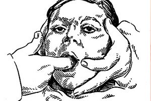
Palpation can reveal hidden pathological conditions or developmental anomalies of the pharynx. For example, this technique can reveal the giant cervical process, which is usually palpated at the border of the posterior edge of the tonsil and the lateral wall of the pharynx; when pressing on the process, the patient may feel pain. Palpation of the cervical processes is performed bimanually: when palpating on the left, the second finger of the left hand is inserted into the oral cavity and the lateral wall of the pharynx is palpated in the above-mentioned area; at the same time, the fingers of the right hand press from the outside at the angle of the lower jaw, trying to penetrate the submandibular fossa in the projection of the exit of the facial nerve.
Palpation of the palatine tonsils can reveal their cicatricial compactions, concretions, as well as pulsating large vessels and aneurysms, which is especially important for planned surgical interventions in this area (removal of tonsils, cervical processes, extended monotonsillectomy for tonsil tumors, opening of peritonsillar abscess, etc.). Using a button probe, penetrate the lacunae, examine their depth, contents, establish the presence of a supratinsilar fossa, etc. By palpation, the condition of the nasopharynx, its walls, as well as the reflex activity of the pharynx and the condition of the lingual tonsil are determined.
Neck examination
Particular attention should be paid to the examination and palpation of the anterior and lateral surfaces of the neck, supraclavicular and jugular fossae. The thyroid gland area, superficial and deep cervical lymph nodes, and projection areas of large cervical vessels are examined. If necessary, a phonendoscope is used to listen to vascular noises in the projection area of the common carotid artery. These noises may occur in pathological conditions of the arteries (aneurysm, stenosis, tumor, etc.) and often simulate tinnitus. They can be differentiated from true tinnitus by compressing the common carotid artery.
Palpation of the neck is performed mainly to determine the condition of the lymph nodes and the thyroid gland. Palpation of the lymph nodes of the neck is performed simultaneously with both hands with the head of the subject slightly tilted forward, starting with the submandibular lymph nodes; then they move on to palpation of the regional lymph nodes for the palatine tonsils, located along the anterior edge of the sternocleidomastoid muscle, then they palpate the deep lymph nodes of the neck along the posterior edge of the said muscle, the supraclavicular and posterior cervical lymph nodes; the latter can be involved in the process in metastatic tumors of the nasopharynx. When palpating the thyroid gland, its size, consistency, and macrostructure are determined. When palpating the jugular fossa and a voluntary swallow of water, it is sometimes possible to detect a lobe of the thyroid gland rising upward, dystopic behind the manubrium of the sternum.
The examination of the pharynx functions is carried out in several directions. First of all, its mobility, symmetry and quality of resonant abilities during phonation are assessed, as well as its swallowing function using a sip of water; in this case, attention is paid to its permeability for liquid. In case of a violation of the swallowing function of the pharynx, the act of swallowing is carried out with effort and forced movements in the neck and trunk, and may be accompanied by pain; in case of paresis of the muscles of the soft palate, liquid gets into the nose, in case of paresis of the muscles that provide protection for the larynx during the act of swallowing, liquid gets into the larynx. As a result of the reverse peristaltic movement of the esophagus, liquid and the contents of the food bolus after a swallow can again return to the oral cavity, etc.
Changes in the timbre of the voice occur with various functional disorders and organic processes in both the innervation and articulatory apparatus. Thus, open nasality occurs with paralysis of the soft palate, its defects, non-closure of the hard palate; closed nasality is observed with obstruction of the nasopharynx ( adenoids, choanal polyp, choanal atresia, nasopharyngeal tumors, etc.). Changes in the timbre of the voice are observed with abscesses and tumors of the pharynx, dysarthria - with defects of the tongue (inability to normally pronounce the sounds t, d, s, e, r) or lips (b, p, v, o, u).
When examining the oral cavity and pharynx, a study of taste sensitivity is carried out at the same time.
Due to the fact that the pharynx occupies a central position in the ENT system anatomically and to a large extent functionally, and its own structure abounds in various and extremely active and vital structures, pathological conditions arising in it manifest themselves not only in known local structural and functional disorders, but also in various organic and functional disorders at a distance. On the other hand, its numerous connections with neighboring organs and regulatory centers of the nervous system, its dependence on the blood supply systems, lymphopoiesis, lymph drainage, etc., often cause the occurrence of certain secondary functional or organic diseases of the pharynx, interpreted as "pharyngeal complications". The wealth of the lymphoid apparatus of the pharynx - a protective instrument often results in various diseases of this apparatus, both local and distant, for example, in the metastasis of purulent or teratogenic emboli. The combination of three most important functions in the pharynx - alimentary, respiratory and immune - significantly diversifies the phenomenology of its diseases, the abundance of which, on the one hand, increases the effectiveness of the probabilistic approach to establishing a specific diagnosis, on the other hand, in a number of cases due to the occurrence of "cross-symptomatology" complicates the differential diagnosis of a number of its diseases.
Situated at the "crossroads" of the respiratory and esophageal tracts, richly supplied with blood and lymphatic vessels, literally saturated with glandular and lymphadenoid tissues, the pharynx is one of the most sensitive organs to various pathogenic factors. Sometimes, when turning to an ENT specialist with a complaint, for example, of minor difficulty in swallowing or choking, the patient (and often the doctor) does not suspect that this symptom may be a manifestation of some progressive disease of the brain or an incipient tumor process, and spontaneously occurring "tonsillitis" may serve as the first sign of a blood disease.
The pharynx is an extremely mobile organ, functioning in strict dependence on the nervous, endocrine and immune regulation of its functions. Malfunctions in any of the above links of the integral regulatory system lead to trophic and functional disorders, entailing secondary, tertiary, etc. pathological changes of an organic nature. The latter, in turn, closing the vicious circle, aggravate the course of the disease, which acquires a systemic character, often turning it into a chronic ongoing process. Based on the above, any, even the most banal disease of the pharynx, should be considered as a condition involving the entire complex of its constituent structures in the pathological process, i.e. as a systemic pathological process requiring an integrated approach, both in diagnostics and in treatment.
It is worth paying attention to one more aspect of the problem of "pharynx disease". It is with diseases of the pharynx and other ENT organs functionally related to it that such a psychosocial state of the patient as his quality of life is subject to significant deterioration. Acute diseases of the pharynx literally "switch off" a person from the social and everyday environment, and chronic diseases, especially those related to specific or professional diseases, can dramatically change the fate of the patient, condemning him to suffering and loneliness.
An important place in the problem under consideration is occupied by diagnostics and treatment of a particular disease. Diagnostics is facilitated by visual and instrumental accessibility of the pharynx, but only if the pathological process is limited by its anatomical limits. However, many diseases of the pharynx have their origins far beyond these limits, and the pharynx acts as a secondary "instance", involved in the pathological process "under duress", and then becoming the organ of the most vivid manifestations. Sometimes a distant focus remains "in the shadows" for a long time, does not manifest itself in any way, and the process in the pharynx is active and vivid. In this case, detection of the primary source is a difficult task, and only a systematic approach to any pathological process, including the study of all possible variants of its causes, increases the likelihood of making the most complete diagnosis, which includes all components of this concept: etiology, pathogenesis and pathoanatomical changes.
Treatment of pharyngeal diseases has its own characteristics. It includes non-surgical, "semi-surgical" (without removing any anatomical structures of the pharynx or opening abscesses) and surgical (adenotomy, tonsillectomy, opening of a retropharyngeal abscess, plastic surgery, oncosurgical interventions). Non-surgical treatment of the pharynx includes local and general use of numerous medicinal products of both herbal and synthetic origin, as well as a number of physiotherapeutic techniques. Local treatment includes compresses, rinsing, inhalations, aerosol and lubricating applications, washing of the lacunae of the palatine tonsils, nasal installations. Physiotherapeutic methods include faradization of the pharynx for various neurogenic diseases, ultraviolet irradiation, for example, for tuberculosis or pharyngeal scleroma, radiation therapy for oncological diseases of the pharynx, etc. Semi-surgical interventions include galvanocautery of the palatine tonsils, dissection of the lacunae, etc. A detailed description of treatment methods is given in the description of specific pharyngeal diseases.
Where does it hurt?
How to examine?