
All iLive content is medically reviewed or fact checked to ensure as much factual accuracy as possible.
We have strict sourcing guidelines and only link to reputable media sites, academic research institutions and, whenever possible, medically peer reviewed studies. Note that the numbers in parentheses ([1], [2], etc.) are clickable links to these studies.
If you feel that any of our content is inaccurate, out-of-date, or otherwise questionable, please select it and press Ctrl + Enter.
Skin angioma
Medical expert of the article

Last reviewed: 05.07.2025
Dermatological pathology – skin angioma – is a localized vascular anomaly in the form of tumor-like growths of deformed capillaries or venules located in the dermis and subcutaneous tissue. These formations can be either protruding above the skin surface or completely flat, often called birthmarks.
Angiomas are one of the most common skin defects. ICD 10 code – class XII (diseases of the skin and subcutaneous tissue), L98.
 [
[ Causes of skin angioma
Today, in dermatology it is customary to distinguish the following forms of skin angioma:
- red birthmark or angioma (cherry angioma, Campbell de Morgan spots);
- port wine stains (or nevus flammeus);
- blue and purple formations or venous lakes;
- spider angioma (arachnoid nevus);
- cavernous angiomas are vascular tumors in the subcutaneous tissue.
Pathogenesis is defined as a congenital defect of the skin and subcutaneous tissue vessels. According to statistics from foreign specialists, this defect is present in 0.7-1.8% of newborns and in 10-15% of premature babies and infants with initially low weight.
Some dermatologists associate the causes of skin angiomas with degenerative changes in the collagen fibers surrounding these blood vessels, which deprives them of the necessary structural support and leads to dilation (i.e. expansion). Another point of view on the pathogenesis of angiomas: the growth of blood vessels in the skin is caused by the proliferation of endothelial cells lining the inner walls of the vessels. The appearance of port-wine stains - diffuse capillary lesions of the skin - is considered to be the result of a violation of the local innervation of the capillary network. However, all these pathological processes have genetic causes. Although skin angiomas in the form of small cherry or red moles (Campbell de Morgan spots) appear in people after 30-40 years (increasing in size and number), as well as after 60 (senile angioma or hemangioma).
There is still no unified classification of skin angiomas and there is a clear terminological inconsistency, which introduces a lot of confusion in the description and diagnosis of these anomalies. Skin angiomas - as a type of congenital vascular pathologies, have a number of names: vascular malformations (defects) of the skin, capillary angiodysplasia, hemangiomas (which may not be congenital and occur at any age), vascular nevi (although nevi are associated with the production of the skin pigment melanin).
Experts from the American National Skin Care Institute differentiate congenital vascular malformations depending on the type of vessels involved: CM (capillary malformation), VM (venous), CVM (capillary-venous), CLM (capillary-lymphatic), LVM (lymphatic-venous), CLVM (capillary-venous-lymphatic malformation), etc.
Complications of skin angiomas may arise from traumatic impact on them, which is fraught with bleeding. The possibility of formation of capillary microthrombi and development of inflammation in the form of purulent granuloma is also not excluded. In addition, it should be noted that the so-called port wine spots of especially large sizes, located on the face, can be with vascular tumors of the pia mater of the brain and indicate a severe congenital pathology of Sturge-Weber-Krabbe syndrome (total damage to the central nervous system with impaired physical and mental development).
Skin angiomas do not pose a cancer risk, and their prognosis is favorable in the vast majority of patients. However, in very rare cases, skin angiosarcoma or malignant hemangioendothelioma are possible.
Symptoms of skin angioma
The first signs of angioma on the skin of various parts of the body in the form of a cherry or red mole - are detected visually at the birth of a child or in an adult. They can be flat or have the shape of a hemisphere, do not disappear when pressed, do not cause discomfort and do not cause any sensations.
It is also easy to recognize a port-wine stain (a type of capillary malformation): it is already present in a newborn; never rises above the skin; has all shades of red and pink, a wide variety of sizes and shapes (with unclear boundaries); is localized on the face or head. These spots can also grow as children grow and acquire a rich purple color. They account for over 10% of vascular malformations.
Diffuse capillary angiomas of the skin, salmon-colored (yellow-pink) and called "stork marks," are located in infants on the scalp, neck, forehead, or eyelids. They regress and disappear on their own over time.
Symptoms of skin angioma in the form of a venous lake (a type of venous malformation) are tumor-like papules of various shapes, bluish, red or purple in color, localized on the lips or eyelids (in children); in old age (65 years and older), they appear on the ears and are more common in men.
Spider angioma of the skin (with a red papule in the center and visible capillaries diverging in different directions) is often defined by dermatologists as a stellate angioma, vascular spider or telangiectasia. They account for up to 40% of all vascular malformations. This angioma is localized along the superior vena cava on the face, neck, hands and forearms, on the upper chest in 10-15% of healthy adults and children. Spider angioma is prone to regression: it can gradually decrease in size, fade, and eventually disappear completely and appear only in the cold or at elevated body temperature.
Such "spiders" can appear in pregnant women, as well as in women using hormonal contraceptives, which may be due to increased estrogen levels. As experts note, the presence of more than three spider angiomas on the skin may be a sign of liver pathology (a third of patients with liver cirrhosis have such "marks" on the skin), and also indicates the likelihood of varicose veins of the esophagus.
The symptoms of a cavernous skin angioma, which is most often called a hemangioma, are the presence of a bluish or purple node in the epidermis that has an uneven surface; when palpated, the formation is hot, and when pressed, it becomes paler. It has a tendency to grow.
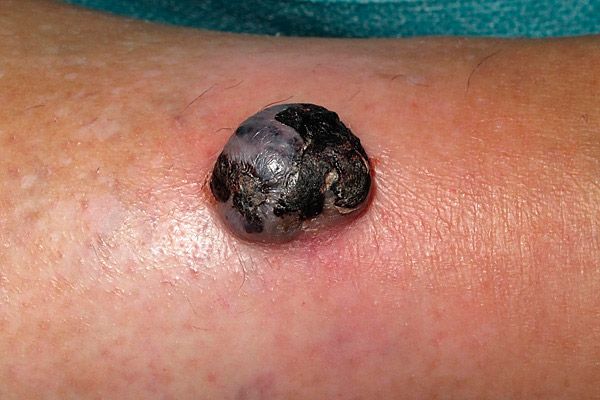
Angiomas of the cavernous type include a congenital red birthmark, called a "strawberry" nevus or nevus vascularis. The first signs may appear several weeks after the birth of the child in the form of a red spot (on the face, head, back and chest). The formation grows rapidly (sometimes up to several centimeters) until about one year of age, and at this stage it looks like a bright red tumor. Then growth stops, and after about a year, a slow regression begins with a change in color to bluish-gray. In 50% of cases, such a birthmark disappears by the age of 5, in 90% by 9; a whitish scar may form in its place. But a large strawberry nevus has negative consequences in the form of an increase in the level of platelets in the blood, which can lead to heart failure.
Diagnosis of skin angioma
Skin angioma is usually diagnosed during a patient examination by a dermatologist, including using a dermatoscope. And for most skin pathologies of this type, there is no need for clinical studies. So, tests are usually not required.
However, when a pathological change in the underlying tissue is detected, instrumental diagnostics (ultrasound scanning) of the vascular formation is necessary.
In more complex cases, when the color and size of a mole or birthmark change, which are not typical for a specific type of angioma, or when the formation bleeds, a full examination is carried out with all tests. If the diagnosis is uncertain (for example, if nodular melanoma or basal carcinoma is suspected), differential diagnostics are carried out using a biopsy and histological examination of the formation tissue, angiography, computed tomography (CT) or magnetic resonance imaging (MRI).
What do need to examine?
How to examine?
Who to contact?
Treatment of skin angioma
Dermatological angioma in most clinical cases does not require treatment; moreover, with the antenatal nature of the pathology, treatment of skin angioma is very problematic. A typical case involves making a diagnosis, explaining to the patient (or parents of children with various birthmarks), the causes and clinical characteristics of the vascular formation and subsequent monitoring of its condition.
Surgical treatment of skin angiomas is performed if the patient is concerned about their appearance, the formations cause discomfort or are located in an area that is easy to touch, which can lead to bleeding. This treatment involves removing them using:
- pulsed laser (PDL);
- cauterization with high-frequency electric current (diathermocoagulation or electrocauterization);
- fulguration (non-contact plasma cauterization);
- cryodestruction (freezing of the protruding nodular angioma with liquid nitrogen);
- punctures and injections of sclerosing agents (alcohol);
- surgical excision.
For drug therapy of cutaneous angiomas, the following medications are used:
- systemic corticosteroids (injections of Prednisolone, Hydrocortisone, etc. into the lesion help stop the growth and accelerate the regression of the angioma);
- interferon α-2a or α-2b (intramuscular administration reduces the proliferation of skin angioma);
- Angiogenesis-inhibiting drugs block vascular endothelial growth factor (VEGF) and are used in pathological neovascularization.
Dermatologists categorically do not recommend self-medication of congenital skin pathologies and, even more so, “testing” folk treatment of skin angioma on children.
The herbal treatment recommended for getting rid of birthmarks has not undergone any tests or clinical trials, so you should not smear a red birthmark, nevus vascularis, with celandine or aloe juice, or a wine birthmark with wormwood decoction. Birch firewood ash or a mixture of apple cider vinegar with honey and black pepper are unlikely to help here.
Tea tree oil, which is quite effective against fungal infections of the skin and nails, is also powerless. In addition, it can cause an allergic reaction.
Skin angioma can have different localizations and often causes some discomfort in cosmetic terms. But this does not affect the general state of health, and prevention of this pathology is impossible, since there are no ways to prevent its occurrence. Go to the doctor, show your child to a specialist and follow medical recommendations.