
All iLive content is medically reviewed or fact checked to ensure as much factual accuracy as possible.
We have strict sourcing guidelines and only link to reputable media sites, academic research institutions and, whenever possible, medically peer reviewed studies. Note that the numbers in parentheses ([1], [2], etc.) are clickable links to these studies.
If you feel that any of our content is inaccurate, out-of-date, or otherwise questionable, please select it and press Ctrl + Enter.
Pineal gland cyst of the brain in adults and children
Medical expert of the article

Last reviewed: 12.07.2025
A pineal gland cyst is a vesicular cavity filled with fluid, namely glandular secretion. Such a cavity is not tumor-like in nature and, as a rule, is not prone to enlargement and progression. But this is not always the case: depending on the size and location, focal symptoms may appear. The diagnosis is established based on the results of MRI or neurosonography (for small children). Treatment, depending on the situation, is either not required or consists of surgery, which is relevant in the development of complications or progressive enlargement of the neoplasm.
Is a pineal gland cyst dangerous?
The human brain is a complex and unique structure. Scientists have been actively studying this organ for many years, but even today many of its areas and functions are considered a mystery to science. The pineal gland, or epiphysis, remains the least studied structure.
The activity of the pineal gland determines the change of rhythms in the human body, such as sleep and wakefulness. In addition, the pineal gland is responsible for the processes of puberty, controls behavioral characteristics, affects homeostasis (for example, regulates the functionality of the cardiovascular system). In general, the main functional areas of the pineal gland are known, but scientists do not yet know many details of these processes.
As for pathologies of the pineal gland, they can be represented by hemorrhages, parasitic diseases and neoplasms of various nature. A pineal gland cyst is a non-tumor formation that develops in one of the lobes. In the overwhelming majority of patients, such neoplasms are small (up to 10-12 mm in size) and do not tend to increase (grow).
Most doctors agree that in the absence of any clinical manifestations directly related to the cystic change (and not to any other pathology), there is no need for global diagnostics and treatment. However, repeated monitoring and determination of the cause of the disorder are necessary, since there are still cases of cystic growth, compression of adjacent structures by it, provoking corresponding somatic and neurological disorders. [ 1 ]
Epidemiology
According to statistics, cystic transformation of the pineal gland is found in approximately 6% of healthy people. In the group of patients who have been diagnosed with such neoplasms, people suffering from periodic migraine pains are found with a higher frequency. For example, in a study of fifty patients diagnosed with a pineal gland cyst, half of the participants complained of migraines (compared to 25% of another group of people who did not have such cystic formations).
Pineal tumors are rare and account for 1% of all intracranial tumors in adults. However, in children they account for up to 8%. Due to the diversity of tumors in this area, the characteristics and epidemiology vary greatly. I will describe each according to the 2016 WHO classification. [ 2 ]
The dynamic results of magnetic resonance imaging were also studied in more than 150 patients with pineal gland cysts. The average age of the participants was 40 years (from 25 to 55 years). The dynamics were studied for a period from six months to 13 years. It was found that during this period, there was virtually no growth of neoplasms, no disorders or deviations. A slight increase in size was noted in only four people, while in 23 cases, the cysts, on the contrary, decreased. Based on this information, the scientists concluded that asymptomatic pineal gland cysts in adults do not require regular diagnostics and neurosurgical consultation. A control MRI procedure one year after the detection of the disorder is sufficient: in the absence of growth and pathological symptoms, further observation is unnecessary. The widespread use of MRI increases the frequency of detection of pineal gland (PE) cysts in clinical neurology. In adults, the prevalence of cysts is 1.1–4.3%. [ 3 ]
During dynamic observation, none of the patients developed any complications related to the neoplasms.
Pineal gland cysts are most often diagnosed:
- in patients aged 20 to 30 years;
- in female patients (approximately three times more often than in men).
In the vast majority of cases, the pathology is asymptomatic and is discovered by chance during an MRI or CT scan of the brain.
Causes pineal cysts
The reasons for the appearance of a pineal gland cyst have not been fully disclosed by scientists. It is known that most often it is a congenital neoplasm or is provoked by a hormonal imbalance disorder. Blockage of the gland's outlet ducts and echinococcal infection can also be the reasons.
During MRI, congenital blockage is visualized, symptoms of impaired fluid drainage are noticeable, which is caused by excessive viscosity of the secretion or tortuosity of the duct. Such a violation rarely poses a danger to the health and life of patients, it does not have a tendency to grow and become malignant.
Parasitic invasion can cause the formation of numerous or large pineal gland cysts. Defective structures are formed by infection with echinococcus, although such pathology is found relatively rarely. Echinococcal cysts develop mainly in people who are engaged in agriculture and raise livestock.
The causes of congenital cyst development have not been fully established. Often the problem is provoked by pregnancy pathologies, drug, alcohol or nicotine addiction of the mother. In such conditions, the future child develops against the background of existing intrauterine hypoxia and intoxication, which has an extremely unfavorable effect on the state of brain structures. Chronic pathologies of the mother, which are in the stage of decompensation, can also be causes.
Risk factors
The main factors influencing the occurrence of a pineal gland cyst include several points. First: the neoplasm can form due to blockage or stenosis of the gland's excretory ducts. This can happen:
- after traumatic brain injury;
- in neuroinfections;
- in autoimmune processes;
- in case of hormonal imbalance;
- in cerebrovascular pathologies.
The second factor is the entry of echinococcus into the body. When penetrating the tissues of the epiphysis, this parasite forms a capsule, which becomes the cystic formation. This type of disorder is detected relatively rarely, but is characterized by special risks.
The third factor is excessive blood supply to the pineal gland, which can lead to hemorrhage. [ 4 ]
As for congenital cystic neoplasms, they are most often detected:
- in children with other intrauterine pathologies;
- in case of diagnosed fetal hypoxia or trauma during labor;
- in infants with postnatal infectious diseases.
Pathogenesis
What does a pineal gland cyst consist of? Its walls are made up of three layers:
- an internal layer of fibrillar glial tissue, often with hemosiderin particles;
- the middle layer is the parenchyma of the epiphysis, which may or may not contain areas of calcification;
- a thin outer layer of fibrous (connective) tissue.
In many cases, the formation of pineal gland cysts is caused by hormonal changes, as such neoplasms are often found in young female patients. Such pathological elements initially actively increase and then subside. In male patients, the condition of the cysts is more stable: intensive growth is usually absent.
The cystic contents are represented by a protein substance that differs from the cerebrospinal fluid on tomographic images. Blood may be present.
Cystic walls tend to actively accumulate contrast. [ 5 ]
With active growth of the neoplasm, the flow of cerebrospinal fluid may be disrupted as a result of blockage (occlusion) of the cerebrospinal fluid-conducting channels, which leads to the development of hydrocephalus.
Symptoms pineal cysts
The overwhelming majority of detected pineal gland cysts are small in size (less than 10 mm in eight out of ten patients), so they do not manifest themselves clinically. If pathological symptoms do appear, then most often this occurs in women over 35 years of age.
Cystic formations of significant dimensions can exert mechanical pressure on the plate of the quadrigeminal body, which entails compression of the superior colliculus and the development of spinal midbrain syndrome (vertical gaze palsy). If pressure is exerted on the Sylvian canal, which is located in the area of the third and fourth ventricles, obstructive hydrocephalus can develop.
If intraosseous hemorrhage occurs, the formation also increases in size: such a pathology is called apoplexy of the pineal gland cyst. [ 6 ]
The following symptoms may appear:
- headaches;
- visual disturbances;
- loss of the ability to move one's gaze up and down;
- lack of coordination of muscle movements in the absence of muscle weakness (ataxia);
- emotional instability;
- mental disorders;
- dizziness, nausea;
- hormonal status disorders (premature puberty, secondary form of Parkinsonism, etc.).
First signs
The first signs of a disorder in a pineal gland cyst may appear only when the formation continues to grow and begins to press on nearby brain structures and blood vessels.
Symptoms in such a situation may include the following manifestations:
- Headaches, prolonged, frequent, of unknown origin, independent of general well-being, weather conditions, etc.
- Dizziness and nausea, constant or paroxysmal, sometimes with vomiting.
- Deterioration of visual and auditory function, blurred vision, double vision.
In severe cases, there may be an unsteady gait, slurred speech, muscle hypertonia, convulsions, deterioration of orientation, loss of reading skills, etc. Similar symptoms may also be associated with increased intracranial pressure, which is also accompanied by drowsiness, inattention, loss of appetite, and swelling of the optic disc.
Acute development of occlusive hydrocephalus, as a complication of the pathological course of cystic neoplasm, manifests itself with signs of increased intracranial pressure. Such signs include:
- headache (especially in the morning);
- nausea with vomiting (after vomiting, the headache may decrease);
- severe drowsiness (precedes a sudden worsening of neurological symptoms);
- congestion of the optic nerve discs (the condition is provoked by an increase in pressure in the subarachnoid space, as well as a change in axoplasmic flow);
- phenomena of axial dislocation of the brain (possibly depression of consciousness up to a deep comatose state, oculomotor disturbances are detected, sometimes a forced position of the head is noted).
With a slow increase in hydrocephalus (chronic course), a triad of signs attracts attention:
- development of dementia;
- disturbance of voluntary movement when walking (apraxia), or paresis of the lower limbs;
- urinary incontinence (the latest and most variable symptom).
Patients become drowsy, inert, and lack initiative. Short-term memory suffers (especially numerical memory). Speech is monosyllabic, often inappropriate. [ 7 ]
Pineal cyst of the pineal gland
The pineal zone is a complex anatomical area that includes the pineal gland, adjacent brain structures, spinal spaces, and vascular network. The pineal gland is located behind the third ventricle, in front of and below it is the posterior cerebral commissure, in front and above is the commissure of the ligaments, below is the quadrigeminal plate and aqueduct, and slightly above and behind is the splenium of the corpus callosum. Directly behind the gland is the quadrigeminal cistern, which forms the cavity of the intermediate velum, which runs above the pineal gland and goes in front below the fornix.
The cyst, which is called pineal, in most cases is not large in size and does not manifest itself clinically. The neoplasm occurs in the epiphysis, without disrupting its function. Only in rare cases, with active growth, can it block the entrance to the cerebral aqueduct, preventing the circulation of cerebrospinal fluid and causing the development of occlusive hydrocephalus.
Pineal gland cyst of the brain in adults
The causes of pineal gland cyst development in adults are still unclear. Scientists voice several theories that could explain the origin of the disorder.
One of these theories suggests the formation of a pathological element due to ischemic or degenerative processes in the glial layer. Some specialists believe that cystic formations are a consequence of the necrosis of the pineal parenchyma. However, the cause of such necrotic processes has not yet been clarified. Other theories of scientists are based on the influence of hemorrhages, hormonal changes, etc. Many such neoplasms are congenital in nature, they are simply discovered by chance at an older age.
The overwhelming majority of such cysts (more than 80%) are small in size - their diameter does not exceed 10 mm. These neoplasms are mostly asymptomatic. Neurological symptoms may appear when such sizes reach 15 millimeters or more.
Cysts accompanied by vivid symptoms are rare. In this regard, specialists do not have extensive information on this issue. As a rule, the very appearance of symptoms and their nature reflect the impact of the neoplasm on nearby structures: the midbrain, internal venous vessels, the vein of Galen, and the optic thalamus. Since the space in this area is extremely limited, it can be expected that even a few millimeters of additional cystic enlargement can cause the appearance of a symptomatic picture, which is most often represented by headache, oculomotor disorders, signs of increased intracranial pressure or the development of hydrocephalus.
Pineal gland cyst in women
In women, pineal gland cysts are found almost three times more often than in men. Many experts associate this with hormonal features. Studies have shown that many cases of such cystic elements began to develop during the onset of puberty, but over the years, such neoplasms appear less and less often. Thus, it is possible to assume a hormonally dependent nature of the occurrence and growth of pineal gland cysts. Moreover, in women, the development of neoplasms is often associated with such hormonal factors as pregnancy and the menstrual cycle. [ 8 ]
Pregnancy with pineal gland cyst
Pregnancy is not a contraindication for a woman who has a pineal gland cyst that does not manifest itself in any way, is asymptomatic and has no tendency to increase.
If the patient has been diagnosed with hydrocephalus or has undergone cerebrospinal fluid shunting surgery, the situation is somewhat different. Pregnancy in such conditions has quite a few risks of complications - for example, shunt function often malfunctions due to increased intra-abdominal pressure due to the constantly growing uterus.
Since the period of pregnancy affects the functional state of the peritoneal-ventricular shunt, doctors have developed a special therapeutic and obstetric management tactic. During the entire period, up to the postpartum stage, the condition of the expectant mother is carefully monitored, all necessary diagnostic procedures for monitoring are performed. [ 9 ]
Is it possible to give birth with a pineal gland cyst?
In the case of an asymptomatic neoplasm, delivery is carried out in the usual manner, taking into account other existing pathologies.
If a peritoneal-ventricular shunt with normal function is present, natural childbirth with a shortened second stage is recommended. Caesarean section under general anesthesia is indicated in cases of impaired shunt function and increased intracranial pressure.
Magnetic resonance imaging is recommended as a safe and effective method for determining the functionality of the shunt and, in general, for assessing the state of the cerebral ventricular system. If functional occlusion of the shunt is noted, drug therapy is administered, with mandatory bed rest and manual pumping procedures.
If an increase in the size of the cerebral ventricles is detected, a surgical operation is prescribed. If we are talking about pregnancy in the first and second trimesters, the operation is performed as if the woman were not pregnant. During the third trimester, alternative methods can be used - in particular, ventriculoatrial shunting or endoscopic triventriculocisternostomy. These methods help prevent the provocation of premature labor and additional trauma to the uterus.
Pineal gland cyst in a child
When a woman hears the diagnosis of "congenital pineal gland cyst" after examining her child, it causes not just concern, but sometimes fear. Let's say right away that in many cases this condition is not so much a pathology as an individual feature, so it does not pose a danger and does not require treatment.
The formation of such cystic formations may be associated with both infections suffered by a woman during pregnancy, and complicated course of this period, or difficult labor. But most often the cause remains unknown. For most epiphyseal cysts, their further development and especially degeneration into an oncological process is not typical.
In infants under one year of age, the presence of such a cyst can be easily determined by ultrasound diagnostics. Childhood up to one year is the most favorable period for performing such a procedure, when the fontanelle is not yet completely closed.
Neurosonography (ultrasound examination of the brain) is especially recommended for premature babies, as well as newborns who, for one reason or another, are subject to intensive care. Complicated labor, complicated pregnancy, intrauterine or intrapartum fetal hypoxia are also indications for ultrasound diagnostics.
Experts believe that the detection of a pineal cyst in a baby should not cause concern. As a rule, such formations do not cause pathology. However, it is advisable to conduct a repeat study after some time to determine the possible dynamics of the process. Most likely, medical observation may be required for a certain period.
In case of unfavorable dynamics, if the formation increases and the fluid pressure in it increases, there is a possibility of changing the position of the surrounding tissues and their compression. Such a disorder manifests itself with such symptoms as seizures, neurological symptoms. In severe cases, the process can be aggravated by the development of hemorrhagic stroke. If there are indications, such a child will be prescribed surgical intervention by one of the existing methods: this can be microneurosurgical, bypass or endoscopic surgery. [ 10 ]
Pineal gland cyst in a teenager
Magnetic resonance imaging of the brain can be prescribed to school-age children and adolescents if there is a suspicion of developing pathology, to diagnose possible disease states. For example, an MRI is prescribed to an adolescent:
- in case of age-related developmental deviations;
- in case of incomprehensible and sudden behavioral changes;
- for regular dizziness;
- for chronic headaches;
- in case of constant fainting or pre-fainting conditions;
- with increasing deterioration of visual or auditory function;
- during convulsive attacks;
- for neurological symptoms.
In the above situations, diagnostics are mandatory. This allows us to identify not only pathological cysts, but also hemorrhages, hydrocephalus, epilepsy, meningitis and meningoencephalitis, etc.
Why can a congenital cyst form? During brain development, the walls of the third ventricle protrude and grow, forming a diverticulum - it is from this that the pineal gland is subsequently formed. If this formation process is disrupted for some reason, incomplete obliteration may occur, and a cavity appears. A small deviation of this kind is not considered pathological, and treatment is not carried out. [ 11 ]
Psychosomatics
Scientists do not exclude the influence of psychological factors on the appearance and growth of neoplasms in the body. This concerns, among other things, pineal gland cysts. And the point is not that a person thinks about the possibility of getting sick and is afraid of it, but that prolonged and strong negative feelings affect the state of brain cells.
According to research, each patient experienced events that were accompanied by strong resentment, anger or deep disappointment before the onset of any tumor processes in the body. From this we can conclude: the problem can be eliminated by neutralizing the internal imbalance.
It is believed that cystic formation is a concentration of the feeling of hopelessness, despair. The disease starts from the moment when the patient stops believing in his own strength, in his loved ones, and becomes disappointed in humanity as a whole.
According to scientists, the following people most often get sick:
- keeping their feelings to themselves, unable to protect themselves and shield themselves from negativity;
- those who do not love themselves, who consider themselves “flawed”, wrong;
- overly emotional about losses;
- those who do not have established contact with their own parents.
Depression and negative emotions begin to put pressure on the immune defense, suppress it, which adversely affects the state of the entire body, even at the cellular level. The immune system is upset, which entails changes in the structure and functionality of cells.
As a rule, such patterns should be identified by the doctor during a conversation with the patient.
Pineal Gland Cyst and Insomnia
Sleep can be called a state of complete rest in the body, in which the most optimal conditions are observed for a person to rest and recover. In particular, his nervous system should be restored. Muscles relax, all types of sensitivity weaken, reflexes are inhibited. However, with some pathologies occurring in the brain, such relaxation is not observed, insomnia occurs, and the quality of sleep is impaired. [ 12 ]
If the pineal gland cyst is large, it can really have a negative impact on the nervous system and sleep. The following symptoms may be observed:
- difficulty falling asleep;
- shallow sleep, with restlessness and frequent awakenings;
- early morning awakening.
We are not talking about absolute insomnia: the patient, although not getting enough sleep, sleeps at least about 5-5.5 hours a day. Much more often, patients experience drowsiness - especially during the day, regardless of the quality of night sleep.
How does a pineal gland cyst affect immunity?
The human brain is directly connected to its immune system, since there are bilateral functional and anatomical connections between these structures. Therefore, it can be assumed that any pathology of the brain, including a pineal gland cyst, can affect the functionality of the immune system, and vice versa. However, for such an effect to occur, the cyst must be large enough to put pressure on nearby tissues. If these dimensions are insignificant, then the immune system is unlikely to suffer: this is the opinion of doctors.
A cyst is not a tumor, therefore it does not cause suppression of the immune defense, unlike malignant primary and metastatic tumor processes of the brain.
Complications and consequences
There are no serious consequences or complications in the vast majority of patients with pineal gland cysts. The probability of malignant transformation is practically zero.
The intensity of symptoms is directly dependent on the size of the formation: thus, cysts up to 10 mm in diameter almost always proceed without any pathological signs.
Large cysts can cause certain complaints, such as migraine headaches, double vision, impaired coordination, nausea, indigestion, fatigue and drowsiness. If such complaints are present, the patient is prescribed a number of diagnostic tests (MRI, biopsy, complete blood count). The main goal of such diagnostics should be to determine the etiology of the disorder and differentiate it from a malignant tumor. The development of hydrocephalus, a pathology that occurs as a result of the release of cerebrospinal fluid from the subarachnoid space, is also considered a threatening condition. Another rare complication in individual patients may be lethargy.
As a rule, conservative treatment is not able to lead to the pineal cyst resolving. The only exception is the early stage of a parasitic neoplasm.
Surgery is not indicated if the cyst does not increase in size and there are no symptoms. [ 13 ]
With a pronounced size of the cystic formation, hydrocephalus may develop - a complication caused by compression or complete squeezing of the Sylvian aqueduct. Almost half of the patients who were referred for surgical treatment had hydrocephalus, which, in turn, was provoked by intracystic hemorrhage. In addition, there are data on isolated cases of syncope and sudden death, which occurred at the moment of sudden blockage of the entrance to the cerebral aqueduct by the cyst.
With increasing hydrocephalus and development of dislocation syndrome, the patient's consciousness is rapidly depressed, up to a deep comatose state. Oculomotor disorders are observed. Compression processes lead to rapid depression of respiration and cardiovascular activity, which, if no assistance is provided, can lead to the death of the patient.
Diagnostics pineal cysts
The main diagnostic method for determining a pineal gland cyst is magnetic resonance imaging. However, in some cases, doctors have to use other diagnostic methods - for example, if the neoplasm is large and accompanied by complex clinical symptoms, or if there is a need for differential diagnostics.
The primary stage is a consultation with a neurologist, passing tests and trials to check reflexes, the degree of skin sensitivity, to assess motor ability. If the patient notes a visual impairment, then he is recommended to consult an ophthalmologist.
Instrumental diagnostics may include the following technical procedures:
- Electroneurography is a specific type of examination for assessing the speed of electrical impulse conduction along the peripheral nerves. The procedure allows determining the degree of nerve damage, as well as the spread and form of the pathological process. This method requires some preparation of the patient: the day before the diagnosis, you should not take sedatives, smoke, drink alcohol and coffee.
- Computer tomography is a type of X-ray examination that involves layer-by-layer visualization of the required area of the brain. In some cases, it can serve as an analogue of MRI.
- Electromyography is a test of the functional capacity of nerve tissue that helps assess the extent of nerve damage and determine motor neuron dysfunction.
- Echoencephaloscopy is one of the harmless ultrasound methods that allows you to assess the state of the functional and anatomical structures of the brain.
- Spinal tap – performed to remove particles of cerebrospinal fluid and then examine it for the presence of atypical cells.
Laboratory tests include:
- general clinical blood and urinetests;
- blood for tumor markers.
A blood test for a pineal gland cyst is not of decisive importance: it is done primarily to assess the general condition of the body, since its results show signs of inflammation (increased ESR and leukocyte levels) and anemia (decreased hemoglobin levels).
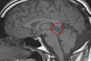
Pineal gland cyst on MRI
The classic version of a pineal cyst is usually small (up to 10 mm) and has one chamber. The diameter of an asymptomatic formation can reach 5-15 mm, and symptomatic cysts sometimes increase even to 45 mm, almost completely replacing the epiphysis.
Every practicing radiologist knows what a pineal gland cyst looks like on MRI: such a neoplasm is voluminous, with liquid contents, with clear configurations. Often (in about every fourth case) peripheral calcifications are present. In many patients, a peripheral contrast accumulation is noted on the image, which has the appearance of a thin and smooth "border". The cyst can change the location of the course of the internal cerebral venous vessels, pushing them upward. [ 14 ]
The following typical signs are observed:
- T1 weighted images:
- typicality of isointense or hypointense signal compared to brain parenchyma;
- in more than half of the cases, the signal is hyperintensified compared to the cerebrospinal fluid;
- signal homogeneity.
- T2 weighted images:
- high signal intensity;
- lower intensity compared to cerebrospinal fluid.
- FLAIR:
- high signal intensity, often not completely suppressed.
- DWI/ADC:
- no diffusion restriction.
- T1-weighted images with contrast enhancement (gadolinium contrast agent):
- more than half of cystic lesions accumulate contrast;
- the contrast accumulates mainly in the form of a thin (less than a couple of millimeters) and even border (full or partial);
- there is a possibility of diffuse contrast enhancement of intracystic fluid with substances containing gadolinium in the late phase (1-1.5 hours), as a result of which the neoplasm acquires a resemblance to a solid volumetric element;
- Sometimes it is possible to detect atypical nodular contrast enhancement or to identify signs of intracystic hemorrhage.
A small pineal gland cyst measuring less than 10-12 mm on MRI or CT has the appearance of a single-chamber fluid formation, with the density of cerebrospinal fluid, or with the same signal activity. Peripheral contrast enhancement is characteristic of the predominant number of cysts, and a band of calcifications (“border”) is observed in approximately every fourth case. [ 15 ]
Single pineal gland cysts are often discovered accidentally during computer or magnetic resonance imaging when diagnosing other pathologies of the brain. In most cases, such formations are not dangerous. However, the doctor must determine not only the location and size of the pathological element, but also whether it corresponds to the neurological symptoms that the patient has.
A multi-chamber pineal gland cyst is typical for echinococcosis of the brain. This pathology can be represented by several varieties:
- solitary type, in which one cyst of fairly large diameter dimensions – up to 6 cm – is formed in the brain;
- racemose type, characterized by the formation of numerous conglomerates of cysts in the form of clusters.
In this situation, MRI becomes the determining diagnostic procedure. It is important to exclude arachnoid cyst, cerebral cysticercosis, epidermoid cyst, intracranial abscess, tumor processes.
An intraparenchymatous pineal cyst is a formation that develops in the parenchyma of the pineal gland and is localized in the posterior parts of the third ventricle (the same pineal region that we mentioned). This neoplasm should be differentiated from pineocytoma, pineoblastoma, and other parenchymatous tumors of the pineal gland. MRI can also detect the disease in this case.
Differential diagnosis
A pineal cyst, especially with nodal contrast enhancement, is virtually indistinguishable from a cystic pineocytoma based on imaging alone. Other neoplasms may develop in the pineal gland, including papillary tumor, germinoma, embryonal cancer, choriocarcinoma, teratoma, arachnoid and epidermoid cysts, vein of Galen aneurysm, and metastatic tumors that have spread to the brain from other sites in the body.
Of course, the cases listed above are rare. However, the results of CT or MRI images should be shown to a qualified neuroradiologist to assess the degree of risk and identify the disease.
Who to contact?
Treatment pineal cysts
For the vast majority of patients, there is no need to treat a pineal cyst. Small neoplasms also do not require further regular follow-up observation, except for one follow-up – 12 months after the first detection of the pathology.
Large elements that are accompanied by pronounced pathological symptoms and pose a danger to the patient are not left without treatment: stereotactic removal of the neoplasm is used, liquid contents are aspirated, communications with cerebrospinal spaces are created, and shunting is performed. In case of relapse of the pineal cyst, radiation therapy is prescribed.
If the formation tends to increase, the patient continues to be observed. If the growth of the pathological element has stopped, then observation continues for another three years.
An absolute indication for surgical treatment is the development of occlusive hydrocephalus and Parinaud's syndrome. Approximately 15% of patients are offered surgery if such painful symptoms as constant dizziness, trembling in the limbs, attacks of nausea and vomiting, disturbances of sensitivity and motor abilities, paroxysmal loss of consciousness appear. Some specialists believe that a pineal gland cyst can provoke a transient obstruction of the Sylvian aqueduct, which manifests itself as headache or clouding of consciousness, especially against the background of a change in body position or with a sharp change in activity.
Although headaches are becoming the most common factor that makes people seek medical help, they may also be the only sign of a disorder associated with a cystic formation. Most doctors (including neurosurgeons) do not associate the presence of a cyst and the occurrence of headaches, provided there is no hydrocephalus. It is indicated that severe headaches can also be explained by central venous hypertension.
In case of pineal gland cyst, physiotherapy treatment is usually not used. [ 16 ]
Medicines
At present, no single treatment strategy has been defined for patients diagnosed with a pineal gland cyst in the absence of hydrocephalus and functional disorders of the midbrain. Most likely, this is due to the lack of complete information about the natural course of the process: many nuances of the origin and development of the cystic formation are unknown, the reasons for its enlargement are not disclosed, and the relationship between the presence of a cyst and the existing clinical picture is not always established. Not all surgeons recommend surgery for patients with non-specific symptoms, and the effectiveness of drug therapy is also controversial. Medicines are prescribed exclusively as symptomatic therapy, depending on clinical indications:
A nonsteroidal anti-inflammatory drug with an analgesic effect. Prescribed for a period of up to 5 days, 1-2 tablets every six hours. A longer course of treatment or exceeding the dosage may negatively affect the digestive system. |
|
Vasobral |
A vasodilator drug that improves blood circulation and metabolism in the brain. Taken orally, with food, 2-4 ml twice a day. The duration of the treatment course is up to 3 months. Possible side effects include nausea and dyspepsia. |
Pikogam |
A nootropic drug with antiplatelet, tranquilizing, psychostimulating and antioxidant effects. Taken orally, regardless of food intake, 0.05 g three times a day for 4-8 weeks. A repeat course is possible in about six months. Possible side effects: allergy, slight nausea, irritability, anxiety. |
Topiramate |
Anticonvulsant drug with antimigraine activity. Treatment begins with the minimum possible dosage, gradually increasing until the desired effect is achieved. The frequency of administration and duration of treatment are determined individually. Possible side effects: loss of appetite, irritability, finger tremors, sleep disturbance, coordination and concentration. |
Analgesic and antipyretic. Take 1-2 tablets orally up to 4 times a day, preferably no longer than three days in a row. Side effects: allergy, nausea, abdominal pain, anemia. |
Herbal treatment
If a patient develops a number of neurological symptoms against the background of a pineal cyst, then in such a situation one should not rely on folk methods of treatment. The fact is that such symptoms, such as memory loss, bilateral visual impairment, muscle weakness, can be a consequence of processes that are dangerous to human life. Therefore, it is better to consult a doctor and follow his recommendations.
Medicinal herbs can be used to relieve headaches, nausea, and to improve cerebral circulation.
- Echinacea extract stimulates metabolic processes in the brain, prevents complications. It is recommended to take Echinacea for a course of at least four weeks.
- Fresh burdock juice has a positive effect on blood circulation, strengthens cerebral vessels, and optimizes nerve conductivity. The juice is squeezed out of leaves that have been previously washed in running water. Drink it on an empty stomach, 1 tbsp in the morning and evening, until you feel a persistent improvement in your health.
- The medicinal herbal collection is prepared from the immortelle plant, chamomile flowers, yarrow, calamus rhizome, calendula, St. John's wort and mint. All ingredients are taken in equal quantities. One tablespoon of the mixture is poured into a thermos with boiling water (400 ml), kept for an hour and a half, then filtered. Take 100 ml 4 times a day half an hour before meals.
It is important to remember that treatment with folk remedies is always long-term: it is important to immediately prepare for several weeks or even months of daily herbal therapy.
Surgical treatment
Since drug treatment for a pineal gland cyst is prescribed only for symptomatic purposes, the only radical method of getting rid of the problem is surgery. A surgeon is sought if the formation continues to grow, signs of hydrocephalus or complications such as bleeding, rupture, or compression of brain structures appear. The neurosurgeon decides which surgical treatment method to choose, depending on the situation. [ 17 ]
If the patient is admitted with impaired consciousness (comatose state or stupor status), he is urgently sent for external ventricular drainage. This procedure helps to relieve the degree of compression of the brain structures and normalize intracranial pressure. Rupture of the cyst or hemorrhage become direct indications for surgical intervention. The patient undergoes cranial trepanation and excision of the neoplasm. [ 18 ]
If there are no complications and no disturbances of consciousness, then the operation is scheduled, using endoscopic access. The main "advantage" of such an intervention is a quick recovery period and relatively little trauma. During endoscopic access, the surgeon makes a burr hole in the cranial bone, through which he aspirates fluid from the cavity. To prevent further accumulation of liquid secretion in the cavity, several holes are made for connection with the cerebrospinal space, or a cystoperitoneal shunting procedure is performed (with the installation of a special shunt). [ 19 ]
The postoperative stage consists of rehabilitation treatment, the appointment of exercise therapy, manual and reflexology. The patient is prescribed medications that improve blood circulation in the brain, as well as decongestants and absorbable drugs.
Pineal Gland Cyst Removal
Today, the use of the latest technologies allows removing brain cysts using endoscopic methods that do not require opening the cranium. Minimally invasive interventions do not entail violations of the integrity of the brain, completely eliminate infection, reduce the risk of complications, and facilitate the recovery period. Unscheduled interventions can be performed on patients in a comatose state or stupor. It is possible to promptly reduce intracranial pressure and eliminate compression of brain structures.
The following surgical techniques can be used:
- Brain shunting is performed to restore cerebrospinal fluid circulation that has been disrupted by a cystic formation. The neurosurgeon uses a drainage tube made of self-absorbing material to ensure fluid flow.
- Endoscopic intervention allows the cyst to be removed through small punctures or transnasally (through the nose). The use of an endoscope with microsurgical instruments and an optical sensor helps to penetrate deep areas to perform the required manipulations.
- Cyst drainage helps to ensure the fluid exits if, for some reason, the patient is contraindicated for surgery.
- Radical resection of the cyst with craniotomy.
The optimal surgical method is selected by the attending physician. Radiosurgical treatment such as gamma knife, cyber knife or radiation therapy is usually not performed. Such methods can only be used to remove a cyst-like tumor of the pineal zone. [ 20 ]
Today, there is a clear trend towards improving the operations performed: this is necessary not only for the effectiveness of treatment, but also to reduce the scale of surgical trauma. Endoscopic surgery is fully suitable for this purpose. [ 21 ]
Currently, clinical institutions use the following technologies for patients with pineal cysts:
- Transnasal (through the nose) endoscopic neurosurgery is practiced in specialized surgical conditions with a variable lighting spectrum, additional monitors for each of the surgeons. The intervention is carried out through the nasal passages using a special neuronavigation system that allows for complete control over the location of the instruments in the surgical field, as well as the position of vital anatomical structures (arterial trunks, optic nerves, etc.). All this helps to perform the operation without further adverse postoperative consequences. Such an intervention is safe for the patient and significantly reduces the duration of inpatient treatment, compared to other surgical techniques.
- Ventricular endoscopy is performed in the area of the cerebral ventricles using special high-tech equipment. During the operation, the neurosurgeon has the opportunity to qualitatively examine the internal brain cavities, make a revision of the cyst and radically remove it. Ventricular endoscopy is prescribed for congenital and acquired cystic neoplasms, for hydrocephalus with occlusion at the level of the third ventricle, cerebral aqueduct, and fourth ventricle.
- Transcranial endoscopy involves the use of a neuroendoscope. The intervention is performed through a mini-access in the form of a cosmetic skin incision with a trepanation window no larger than 20-25 mm. This technology allows for a significant reduction in brain trauma with optimal visualization of the painful area, as well as performing the intervention with minimal blood loss. An additional "plus" is the excellent cosmetic result.
Prevention
Acquired forms of pineal gland cysts are most often the result of inflammatory processes, injuries, vascular and infectious pathologies. Therefore, it becomes clear that only correct and timely treatment of all kinds of diseases and traumatic injuries can become the optimal prevention of the development of cystic formations in the brain. During the treatment of inflammatory, infectious and vascular pathologies, one should not forget about the implementation of resorption and neuroprotective therapy.
To prevent the development of congenital cysts it is necessary:
- correct pregnancy management;
- adequate management of labor;
- prevention of fetal hypoxia;
- prevention of the development of fetoplacental insufficiency;
- conducting explanatory work with expectant mothers and pregnant women regarding the need to lead a healthy lifestyle;
- prevention of intrauterine infection;
- prohibition of pregnant women from taking certain medications;
- special control in case of negative Rh factor in the expectant mother.
Forecast
The overwhelming majority of cases of pineal gland cysts have a favorable prognosis: it has been established that in 70-80% of patients such neoplasms do not increase or even decrease in size throughout life.
Experts note that in the absence of symptoms associated with the presence of a pineal gland cyst, no therapeutic or surgical measures should be taken. Periodically - approximately once every three years - control diagnostics can be performed in the form of computed tomography or magnetic resonance imaging. However, this is not always necessary: in the absence of growth of the formation, it is sufficient to observe patients only in the clinical spectrum. [ 22 ]
Magnetic resonance imaging monitoring is necessary if the cyst has diameter dimensions exceeding 10-12 mm: in such a situation, the pathology should be diagnosed with cystic pineocytoma.
Disability
A pineal gland cyst itself cannot be the basis for establishing a patient's disability. In order for a person to be recognized as disabled and assigned the appropriate disability group, he or she must have persistent (that is, those that do not respond to either conservative or surgical treatment) and obvious functional impairments of the body.
As a rule, minor disorders such as migraines and visual impairment do not become grounds for assigning a disability group.
The patient is considered incapacitated if, as a result of the pineal gland cyst, he develops the following persistent symptoms:
- epileptic seizures;
- movement disorders in the form of para-, hemi- and tetraparesis;
- severe dysfunction of the pelvic organs (for example, urinary and/or fecal incontinence);
- severe disturbances of the vestibular apparatus;
- progressive mental disorders;
- bilateral deterioration (loss) of hearing function, bilateral severe deterioration (loss) of vision.
An assessment of the presence or absence of signs of disability (indications for establishing disability) in a patient is carried out only at the end of the required course of treatment, and not earlier than 4 months from the start of treatment (or not earlier than 4 months after the surgical intervention).
Pineal cyst and the army
In the vast majority of cases, a pineal gland cyst does not cause concern: convulsions, pain, hearing and vision disorders are extremely rare. A cyst is not an oncological disease. Therefore, in order to determine the suitability of a conscript for military service, doctors need to assess the functional capacity of the body, the degree of severity of disorders (if any). For example, they will carefully study all possible emotional, mental, neurological and other clinical deviations.
If a pineal gland cyst is detected in a patient during an MRI, but it does not manifest itself clinically (does not bother), then the conscript will be considered fit for military service due to this pathology - with only some restrictions regarding the type of troops. If the neoplasm demonstrates moderate or severe disorders of the nervous system, the young man has the right to expect exemption from conscription. The corresponding category is assigned if damage to the nervous system is proven.