
All iLive content is medically reviewed or fact checked to ensure as much factual accuracy as possible.
We have strict sourcing guidelines and only link to reputable media sites, academic research institutions and, whenever possible, medically peer reviewed studies. Note that the numbers in parentheses ([1], [2], etc.) are clickable links to these studies.
If you feel that any of our content is inaccurate, out-of-date, or otherwise questionable, please select it and press Ctrl + Enter.
Increased intracranial pressure (intracranial hypertension)
Medical expert of the article

Last reviewed: 04.07.2025
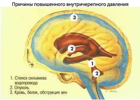
Causes of increased intracranial pressure
Causes of increased intracranial pressure may be the following:
- Occlusion of the ventricular system due to congenital or acquired lesions.
- Volumetric intracranial processes, including hemorrhages.
- Impaired absorption of cerebrospinal fluid by the arachnoid granulations, which may be damaged by diseases such as meningitis, subarachnoid hemorrhage, or brain injury.
- Idiopathic intracranial hypertension (pseudotumor cerebri).
- Diffuse cerebral edema following blunt head trauma.
- Severe systemic hypertension.
- Hypersecretion of cerebrospinal fluid by a tumor of the choroid plexus, which is very rare.
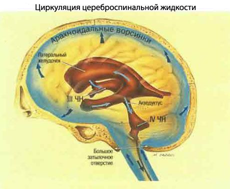
Circulation of cerebrospinal fluid
- Cerebrospinal fluid (CSF) is formed by the choroid plexuses in the ventricles of the brain.
- Leaves the lateral ventricles, entering the third ventricle through the foramen of Monro.
- From the third ventricle, through the Sylvian aqueduct, it enters the fourth ventricle.
- From the fourth ventricle, cerebrospinal fluid (CSF) passes through the foramina of Luschka and Magendie into the subarachnoid space, flows around the spinal cord, and then washes the cerebral hemispheres.
- It is absorbed into the venous drainage system of the brain through the granulations of the arachnoid membrane.
Normal CSF pressure at lumbar puncture is <80 mm H2O in infants, <% mm H2O in children, and <210 mm H2O in adults.
Symptoms of increased intracranial pressure
Symptoms of increased intracranial pressure include a pressing headache, vomiting, and swelling of the optic nerve papilla.
With prolonged increase in intracranial pressure, the level of consciousness decreases, weakened or asymmetrical pupillary response gradually disappears completely, hypertension and bradycardia, loss of consciousness, and death are observed.
Features of increased intracranial pressure in children
- The relatively large head volume and weak neck muscles make the child's brain more susceptible to acceleration-deceleration injuries.
- In children under 2 years of age, brain swelling may be compensated for by expansion of the cranial bones and can be assessed by observing the fontanelles and measuring the head circumference. Skull fractures are less common in them than in adults.
- Soft tissue wounds of the head and intracranial hematomas can cause hypotension due to the relatively large size of the head and small CBV.
- Intracranial hematomas requiring surgical treatment are less common than in adults (20-30% of TBI in children and 50% in adults).
- Cerebral blood flow is higher in children than in adults, and this may provide some "protection" against ischemic damage.
- Neurological outcomes in children are better than in adults with the same GCS score after resuscitation.
Where does it hurt?
What's bothering you?
Hydrocephalus
Hydrocephalus is an enlargement of the ventricles.
Increased intracranial pressure can be associated with two types of hydrocephalus.
Communicating hydrocephalus, in which cerebrospinal fluid passes without difficulty from the ventricular system into the subarachnoid space. The obstruction to the flow of cerebrospinal fluid is located in the basal cisterns or in the subarachnoid space, where absorption by the pacchionian granulations may be impaired.
Noncommunicating hydrocephalus is associated with a disruption of the flow of cerebrospinal fluid in the ventricular system or in the outlet openings of the fourth ventricle. Because of this, cerebrospinal fluid does not reach the subarachnoid space.
 [ 16 ], [ 17 ], [ 18 ], [ 19 ], [ 20 ]
Symptoms of hydrocephalus
Systemic symptoms of hydrocephalus
- Headaches may occur at any time of the day, especially in the morning, which may interrupt sleep. As a rule, pain that increases over 6 weeks leads the patient to see a doctor. Headaches may be generalized or localized and increase with head movements, bending over, or coughing. Patients who have suffered from headaches before may report a change in their nature. Very rarely, headaches may be absent.
- Sudden nausea and vomiting, often severe, may provide some relief from headache. Vomiting may be an independent symptom or may precede headaches by up to a month, especially in patients with fourth ventricular tumors.
- Impairment of consciousness may be mild, with drowsiness and sleepiness. Sudden significant impairment indicates damage to the brainstem with tentorial or cerebellar herniation and requires immediate action.
Visual symptoms of hydrocephalus
- Transient visual disturbances lasting a few seconds are common in patients with occluded disc disease.
- Horizontal diplopia is caused by tension of the abducens nerve over the pyramid. This is a false topical symptom.
- Visual impairment appears later in patients with secondary optic nerve atrophy due to long-standing stagnation of the disc.
Idiopathic intracranial hypertension
Idiopathic intracranial hypertension deserves special mention because it may require ophthalmology. Idiopathic intracranial hypertension is defined as elevated intracranial pressure in the absence of an intracranial mass lesion or ventricular dilation due to hydrocephalus. Although idiopathic intracranial hypertension is not life-threatening, permanent visual impairment due to disk congestion may occur. Ninety percent of patients are obese women of childbearing age, often with amenorrhea. Intracranial hypertension may also be caused by medications, including tetracyclines, nalidixic acid, and iron supplements.
[ 24 ]
Features of idiopathic increased intracranial pressure
- Complaints and symptoms of increased intracranial pressure, as described earlier.
- Lumbar puncture reveals pressure >210 mm H2O. Pressure may also be elevated in obese patients with normal intracranial pressure.
- Neurological studies show normal or small and slit-shaped ventricles.
[ 25 ]
The course of idiopathic increased intracranial pressure
In most patients the course is long, with spontaneous relapses and remissions, in some it may last only a few months. Mortality is low, visual impairment is frequent and sometimes severe.
How to recognize increased intracranial pressure?
- Intracranial pressure greater than 25 mmHg, measured by an intraparenchymal microtransducer or external ventricular drain - lateral ventricular cerebrospinal fluid pressure is the "gold standard" for measuring intracranial pressure.
- Identifiable intracranial pressure wave abnormalities often arise as a result of phasic cerebral vasodilation in response to a fall in cerebral perfusion pressure (CPP) and resolve with an increase in BP.
- plateau ("A") of waves paroxysmally increases to 50-100 mm Hg (usually against the background of initially high pressure) and usually lasts for several minutes (up to 20 min);
- "B" waves are significantly shorter fluctuations, lasting about a minute and reaching 30-35 mm Hg at their peak;
- Abnormal intracranial pressure waves reflect decreased intracranial compliance.
Treatment of increased intracranial pressure
Treatment of increased intracranial pressure has two goals - reducing headaches and preventing blindness.
Regular perimetry is important to detect initial and progressive changes in the visual field.
Treatment of increased intracranial pressure requires the use of the following medications and methods:
- Diuretics such as acetazolamide or thiazides usually reduce headache, but their effect on preservation of visual function is unknown.
- Systemic steroids are often used short-term rather than long-term because of potential complications, especially in obese patients.
- Fenestration of the optic nerve, which involves cutting its meninges, reliably and effectively preserves vision if performed in a timely manner. However, it rarely reduces headaches.
- Lumboperitoneal shunts can be used, but often require surgical revision due to failure.
Emergency treatment of increased intracranial pressure
- Sedation and analgesia to reduce brain metabolic activity and minimize blood pressure fluctuations.
- Mechanical ventilation to maintain PaO2 > 13.5 kPa (100 mmHg) and PaCO2 4.0-4.5 kPa (30-34 mmHg).
- Position with the head end of the table raised by 15-20°, neutral position of the neck, exclude obstruction of the veins of the neck.
- Maintain adequate BP (>60 mmHg), but correct hypertension if SBP >130 mmHg.
- Mannitol 20% (0.5 g/kg) or other osmotic diuretic.
[ 31 ], [ 32 ], [ 33 ], [ 34 ], [ 35 ], [ 36 ]
Further management
- Maintain IVPP > 60 mmHg to ensure adequate brain oxygenation with volume replacement therapy and inotropes/vasopressors.
- Treat BP when it rises above the upper limit of autoregulation (SBP > 60 mmHg) to minimize vasogenic brain swelling using short-acting drugs such as labetalol and esmolol.
- Moderate hyperventilation to PaCO2 4.0-4.5 kPa (30-34 mmHg). Hyperventilation to PaCO2 <4.0 kPa (30 mmHg) is only permissible under conditions of cerebral oxygenation monitoring (e.g., using jugular vein oximetry) - excessive hyperventilation can worsen cerebral ischemia by further reducing the critically low cerebral blood flow.
- Treat hyperthermia.
- Consider moderate induced hypothermia (target 34 CC). Although prospective randomized trials have not shown improved outcomes with this approach, moderate temperature reduction is effective in reducing elevated intracranial pressure.
- Mannitol (0.5 g/kg), usually as a 20% solution.
- Drainage of cerebrospinal fluid through a ventricular catheter is effective in reducing elevated intracranial pressure, but the procedure is invasive and not without risks.
- Bone flap removal (decompressive craniectomy) with dura mater reconstruction is a therapeutic approach for intracranial hypertension refractory to conventional therapy.
[ 37 ]