Penis
Last reviewed: 25.06.2018

All iLive content is medically reviewed or fact checked to ensure as much factual accuracy as possible.
We have strict sourcing guidelines and only link to reputable media sites, academic research institutions and, whenever possible, medically peer reviewed studies. Note that the numbers in parentheses ([1], [2], etc.) are clickable links to these studies.
If you feel that any of our content is inaccurate, out-of-date, or otherwise questionable, please select it and press Ctrl + Enter.
The penis serves to remove urine from the bladder and to release semen into the female genital tract. The penis consists of a free anterior part - the body (corpus penis), which ends with the head (glans penis), which has a slit-like external opening of the male urethra (ostium urethrae externum) at its apex.
Pain around the head of the penis
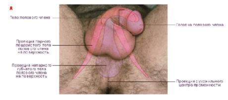
The head of the penis has the widest part, the crown of the head (corona glandis), and the narrowed part, the neck of the head (collum glandis). The back part, the root of the penis (radix penis), is attached to the pubic bones. The upper anterior surface of the body is called the back of the penis (dorsum penis).
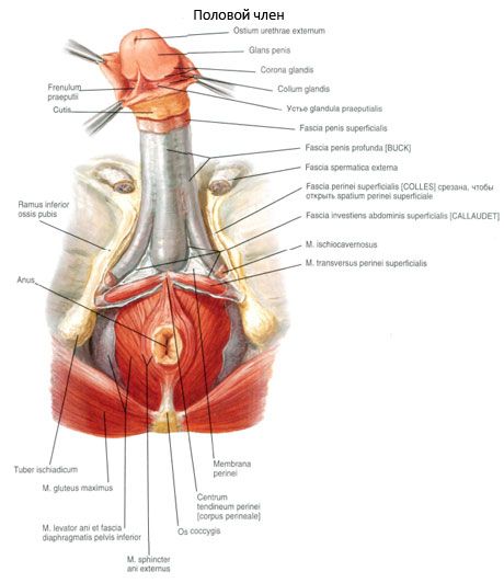
The body of the penis is covered with thin, light, shifting skin, which passes into the skin of the pubis at the top and the skin of the scrotum at the bottom. On the skin of the lower surface of the penis there is a raphe penis, which continues posteriorly onto the skin of the scrotum and perineum. In the anterior part of the body of the penis, the skin forms a well-defined skin fold - the foreskin of the penis (preputium penis), which covers the head, then passes into the skin of the glans penis. The foreskin is attached to the neck of the head. Between the glans penis and the foreskin there is a cavity of the foreskin, which opens in front with an opening that allows the glans penis to pass through when the foreskin is pulled back. On the underside of the glans penis, the foreskin is connected to the glans by the frenulum of the foreskin (frenulum preputii), which almost reaches the edge of the external opening of the urethra. The inner surface of the skin fold, as well as the head, are covered with thin, delicate, translucent skin, different from the skin covering the body of the penis. The skin of the inner layer of the foreskin contains the glands of the foreskin (gll.preputiales).
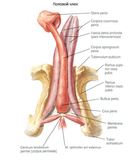
The penis has a paired cavernous body of the penis (corpus cavernosum penis), right and left. They are located next to each other. Under them lies the unpaired spongy body of the penis (corpus spongiosum penis). Each cavernous body is cylindrical in shape. The posterior ends of the cavernous bodies are pointed, diverge to the sides in the form of the legs of the penis (crura penis), which are attached to the lower branches of the pubic bones. The cavernous bodies are fused with each other by their medial surfaces and are covered with a common protein coat of the cavernous bodies (tunica albuginea corporum caver nosorum), which forms the septum of the penis (septum penis) between the cavernous bodies. The spongy body of the penis is expanded in the posterior (proximal) section and forms the bulbus penis (bulbus penis). The anterior (distal) end of the spongy body is sharply thickened and forms the head of the penis. The spongy body is covered by its own protein coat of the spongy body (tunica albuginea corporis spongiosi) and is penetrated throughout its entire length by the urethra, which ends at the head of the penis with an external opening that looks like a vertical slit.
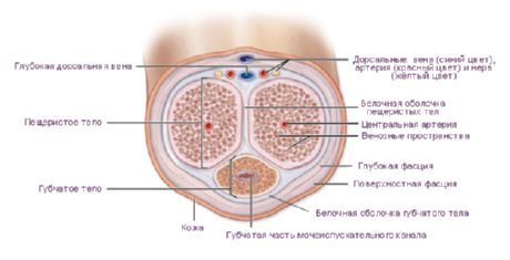
The cavernous and spongy bodies of the penis consist of numerous connective tissue crossbars - trabeculae - branching off from the protein coat, delimiting a system of interconnected cavities (cells) lined with endothelium. When filled with blood, their walls straighten, the cavernous and spongy bodies of the penis swell and become dense (erection of the penis).
The cavernous and spongy bodies of the penis are surrounded by connective tissue plates - the deep and superficial fascia (fascia penis profunda et fascia penis superficialis). On the back of the penis, closer to its root, the fascia are better expressed due to the fact that in this place the tendons of the bulbospongiosus and ischiocavernous muscles pass into them. Outside the superficial fascia there is skin. The penis is also fixed by two suspensory ligaments - superficial and deep. The superficially located suspensory ligament of the penis begins on the lower surface of the fascia of the abdomen, in the area of the white line, and is woven into the superficial fascia of the penis. The deep sling-shaped ligament (lig.fundiforme) has the shape of a triangle, comes from the lower part of the pubic symphysis, divides into two bundles and is woven into the protein tunic of the lateral surfaces of the cavernous bodies.
 [
[ Vessels and nerves of the penis
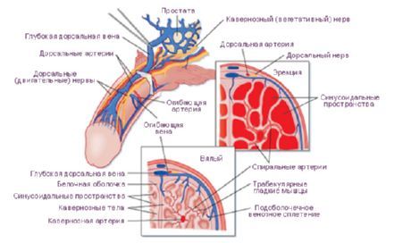
The skin and membranes of the penis are supplied with blood by the anterior scrotal branches from the external genital arteries and by the dorsal artery of the penis from the internal genital artery. The cavernous and spongy bodies of the penis receive blood through the deep artery of the penis and the dorsal artery of the penis, both from the internal genital artery. The arteries of the bulb of the penis enter the bulb of the penis, and the arteries of the urethra (branches of the internal genital artery) enter the spongy body.
Venous blood from the penis flows through the deep dorsal vein of the penis and through the vein of the bulb of the penis into the vesical venous plexus, and also through the deep veins of the penis into the internal pudendal vein.
The lymphatic vessels of the penis drain into the internal iliac and superficial inguinal lymph nodes. The sensory nerve is the dorsal nerve of the penis from the pudendal nerve. Sympathetic fibers come from the inferior hypogastric plexuses, and parasympathetic fibers come from the pelvic visceral nerves.
Last reviewed: 25.06.2018