
All iLive content is medically reviewed or fact checked to ensure as much factual accuracy as possible.
We have strict sourcing guidelines and only link to reputable media sites, academic research institutions and, whenever possible, medically peer reviewed studies. Note that the numbers in parentheses ([1], [2], etc.) are clickable links to these studies.
If you feel that any of our content is inaccurate, out-of-date, or otherwise questionable, please select it and press Ctrl + Enter.
Bladder
Medical expert of the article

Last reviewed: 04.07.2025

The urinary bladder (vesica urinaria) is an unpaired hollow organ that functions as a reservoir for urine, which is excreted from the bladder through the urethra.
The shape and size of the bladder change as it fills with urine. A full bladder has a rounded shape: The capacity of the bladder in an adult is up to 250-500 ml.
The urinary bladder has an anterior superior part, which faces the anterior abdominal wall, and the apex of the bladder (apex vesicae). From the apex of the bladder to the navel there is a fibrous cord - the median umbilical ligament (lig.umbilicale medianum) - a remnant of the embryonic urinary duct (urachus). Without a distinct border, the apex of the bladder passes into the expanding part - the body of the bladder (corpus vesicae). Continuing backwards and downwards, the body of the bladder passes into the bottom of the bladder (fundus vesicae). The lower part of the urinary bladder narrows in a funnel-shaped manner and passes into the urethra. This part is called the neck of the bladder (cervix vesicae).
 [
[ Topography of the urinary bladder
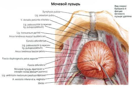
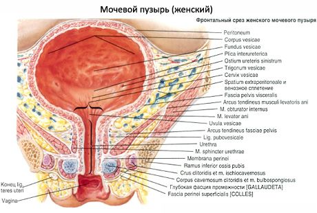
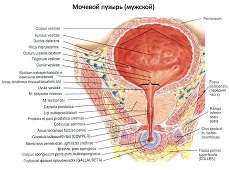
The urinary bladder is located in the cavity of the small pelvis behind the pubic symphysis. Its anterior surface faces the pubic symphysis, from which it is separated by a layer of loose tissue located in the retropubic space. When the urinary bladder is filled with urine, its apex protrudes beyond the pubic symphysis and contacts the anterior abdominal wall. The posterior surface of the urinary bladder in men is adjacent to the rectum, seminal vesicles and ampullae of the vas deferens, and the bottom is adjacent to the prostate gland. In women, the posterior surface of the urinary bladder is in contact with the anterior wall of the cervix and vagina, and the bottom is in contact with the urogenital diaphragm. The lateral surfaces of the urinary bladder in men and women border on the muscle that lifts the anus. The loops of the small intestine are adjacent to the upper part of the urinary bladder in men, and the uterus is adjacent to the upper part of the urinary bladder in women. A full urinary bladder is located mesoperitoneally in relation to the peritoneum, while an empty, collapsed bladder is located retroperitoneally.
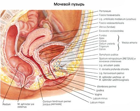
The peritoneum covers the urinary bladder from above, from the sides and from behind, and then in men it passes onto the rectum (rectovesical recess), in women - onto the uterus (vesicouterine recess). The peritoneum covering the urinary bladder is loosely connected to its walls. The urinary bladder is fixed to the walls of the small pelvis and is connected to adjacent organs by fibrous cords. The apex of the bladder is connected to the navel by the median umbilical ligament. The lower part of the urinary bladder is attached to the walls of the small pelvis and adjacent organs by ligaments formed by compacted connective tissue bundles and fibers of the so-called pelvic fascia. In men, there is a puboprostatic ligament (lig.puboprostaticum), and in women - a pubovesical ligament (lig.pubovesicale). In addition to the ligaments, the bladder is also strengthened by muscle bundles that form the pubovesical muscle (m.pubovesicalis) and the rectovesical muscle (m.rectovesicalis). The latter is present only in men. In both men and women, the bladder is to some extent fixed by the initial part of the urethra and the terminal sections of the ureters, as well as the prostate gland in men and the urogenital diaphragm in women.
Structure of the urinary bladder
The walls of the urinary bladder (in men and women) consist of a mucous membrane, submucosa, muscular membrane and adventitia, and in places covered by the peritoneum there is a serous membrane. When the urinary bladder is full, the walls are stretched, thin (2-3 mm). After emptying, the bladder decreases in size, its wall contracts due to the muscular membrane and reaches a thickness of 12-15 mm.
The mucous membrane (tunica mucosa) lines the bladder from the inside and forms folds when the bladder is empty. When the bladder is filled with urine, the folds of the mucous membrane are completely straightened. The epithelial cells (transitional) covering the mucous membrane are rounded when the bladder is empty, and when it is filled and the walls are stretched, they are flattened and thinned. The epithelial cells are connected to each other by tight contacts. In the thickness of the proper plate of the mucous membrane there are alveolar-tubular glands, nerve fibers, vessels and lymphoid formations. The mucous membrane is pinkish, mobile, easily gathers into folds, with the exception of a small area in the area of the bottom of the bladder - the triangle of the bladder (trigonum vesicae), where it is tightly fused with the muscular membrane. In the anterior part of the bottom of the bladder (at the apex of the triangle) on the mucous membrane there is an internal opening of the urethra, and in each corner of the triangle (at the ends of the posterior border) there is an opening of the ureter (right and left; ostium ureteris, dextrum et sinistrum). Along the base (posterior border) of the bladder triangle runs the interureteral fold (plica interureterica).
The submucosa (tela submucosa) is well developed in the wall of the bladder. Thanks to it, the mucous membrane can gather into folds. In the area of the triangle of the bladder, the submucosa is absent. Outside of it, in the wall of the bladder, there is a muscular membrane (tunica muscularis), consisting of three indistinctly delimited layers formed by smooth muscle tissue. The outer and inner layers have a predominantly longitudinal direction, and the middle, most developed, is circular. In the area of the neck of the bladder and the internal opening of the urethra, the middle circular layer is most well expressed. At the beginning of the urethra, the sphincter of the bladder (m.sphincter vesicae) is formed from this layer. When the muscular membrane of the bladder contracts and the sphincter opens at the same time, the volume of the organ decreases and urine is expelled through the urethra. In connection with this function of the muscular membrane of the bladder, it is called the muscle that pushes out urine (m.detrusor vesicae).
Vessels and nerves of the bladder
The superior vesical arteries, branches of the right and left umbilical arteries, approach the apex and body of the bladder. The lateral walls and bottom of the bladder are supplied with blood by branches of the inferior vesical arteries (branches of the internal iliac arteries).
Venous blood from the wall of the urinary bladder flows into the venous plexus of the urinary bladder, and also through the vesical veins directly into the internal iliac veins. The lymphatic vessels of the urinary bladder flow into the internal iliac lymph nodes. The urinary bladder receives sympathetic innervation from the inferior hypogastric plexus, parasympathetic innervation through the pelvic visceral nerves, and sensory innervation from the sacral plexus (from the genital nerves).
X-ray anatomy of the bladder
When filled with a contrast mass, the urinary bladder on the radiograph (in the anteroposterior projection) has the shape of a disk with smooth contours. In the lateral projection on the radiograph, the urinary bladder takes the shape of an irregular triangle. Cystoscopy (examination of the mucous membrane) is also used to examine the urinary bladder. This method allows determining the condition, color, relief of the mucous membrane, ureter openings and the flow of urine into the urinary bladder.
The urinary bladder of newborns is spindle-shaped, in children of the first years of life it is pear-shaped. During the second childhood (8-12 years) the urinary bladder is ovoid, and in adolescents it has a shape typical of an adult. The capacity of the urinary bladder of newborns is 50-80 cm 3, by 5 years - 180 ml of urine, and in children over 12 years old it is 250 ml. In a newborn, the bottom of the urinary bladder is not formed, the triangle of the urinary bladder is located frontally and is part of the posterior wall of the bladder. The circular muscle layer in the wall of the bladder is poorly developed, the mucous membrane is well developed, the folds are pronounced.
The topography of the bladder in a newborn is such that its apex reaches half the distance between the navel and the pubic symphysis, so the bladder in girls at this age does not come into contact with the vagina, and in boys - with the rectum. The anterior wall of the bladder is located outside the peritoneum, which covers only its posterior wall. At the age of 1-3 years, the bottom of the bladder is located at the level of the upper edge of the pubic symphysis. In adolescents, the bottom of the bladder is at the level of the middle, and in adolescence - at the level of the lower edge of the pubic symphysis. Subsequently, the bottom of the bladder descends depending on the state of the muscles of the urogenital diaphragm.
Использованная литература